

7 Nature Driven Magnetic Nanoarchitectures
161
1 μm
(a)
Anoxic
Oxic
O 2
Water
Column
Sediment
(b)
Oxic-anoxic
transiƟon
zone (OATZ)
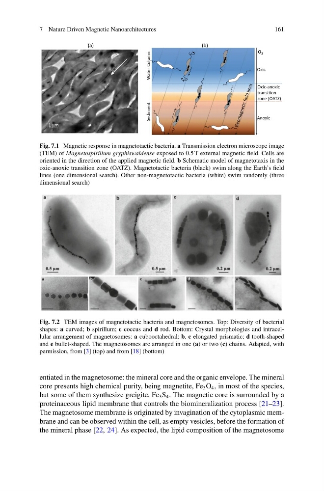
Fig. 7.1 Magnetic response in magnetotactic bacteria. a Transmission electron microscope image
(TEM) of Magnetospirillum gryphiswaldense exposed to 0.5 T external magnetic field. Cells are
oriented in the direction of the applied magnetic field. b Schematic model of magnetotaxis in the
oxic-anoxic transition zone (OATZ). Magnetotactic bacteria (black) swim along the Earth’s field
lines (one dimensional search). Other non-magnetotactic bacteria (white) swim randomly (three
dimensional search)
Fig. 7.2 TEM images of magnetotactic bacteria and magnetosomes. Top: Diversity of bacterial
shapes: a curved; b spirillum; c coccus and d rod. Bottom: Crystal morphologies and intracellular arrangement of magnetosomes: a cubooctahedral; b, c elongated prismatic; d tooth-shaped
and e bullet-shaped. The magnetosomes are arranged in one (a) or two (c) chains. Adapted, with
permission, from [3] (top) and from [18] (bottom)
entiated in the magnetosome: the mineral core and the organic envelope. The mineral
core presents high chemical purity, being magnetite, Fe 3 O 4 , in most of the species,
but some of them synthesize greigite, Fe 3 S 4 . The magnetic core is surrounded by a
proteinaceous lipid membrane that controls the biomineralization process [21–23].
The magnetosome membrane is originated by invagination of the cytoplasmic membrane and can be observed within the cell, as empty vesicles, before the formation of
the mineral phase [22, 24]. As expected, the lipid composition of the magnetosome
161
1 μm
(a)
Anoxic
Oxic
O 2
Water
Column
Sediment
(b)
Oxic-anoxic
transiƟon
zone (OATZ)
Fig. 7.1 Magnetic response in magnetotactic bacteria. a Transmission electron microscope image
(TEM) of Magnetospirillum gryphiswaldense exposed to 0.5 T external magnetic field. Cells are
oriented in the direction of the applied magnetic field. b Schematic model of magnetotaxis in the
oxic-anoxic transition zone (OATZ). Magnetotactic bacteria (black) swim along the Earth’s field
lines (one dimensional search). Other non-magnetotactic bacteria (white) swim randomly (three
dimensional search)
Fig. 7.2 TEM images of magnetotactic bacteria and magnetosomes. Top: Diversity of bacterial
shapes: a curved; b spirillum; c coccus and d rod. Bottom: Crystal morphologies and intracellular arrangement of magnetosomes: a cubooctahedral; b, c elongated prismatic; d tooth-shaped
and e bullet-shaped. The magnetosomes are arranged in one (a) or two (c) chains. Adapted, with
permission, from [3] (top) and from [18] (bottom)
entiated in the magnetosome: the mineral core and the organic envelope. The mineral
core presents high chemical purity, being magnetite, Fe 3 O 4 , in most of the species,
but some of them synthesize greigite, Fe 3 S 4 . The magnetic core is surrounded by a
proteinaceous lipid membrane that controls the biomineralization process [21–23].
The magnetosome membrane is originated by invagination of the cytoplasmic membrane and can be observed within the cell, as empty vesicles, before the formation of
the mineral phase [22, 24]. As expected, the lipid composition of the magnetosome