

Compared to oxidases, the O 2 independence of DHs brings a remarkable benefit
to electrochemical biosensors. In the case of glucose biosensing, there was much
hope that glucose dehydrogenase (GDH) would replace GOx, as has it happened
indeed. Due to the vital importance of controlling blood glucose levels in diabetes
patients, glucose meters account for the largest share of the global point-of-care
diagnostic market [150]. Therefore, the development of new glucose biosensors
involving GDH has been the object of great attention. As a result, modern blood
glucose meters comprise electrochemical cells containing PQQ-GDH, NAD-GDH,
FAD-GDH, and a redox mediator [151]. As mentioned above, the main advantage
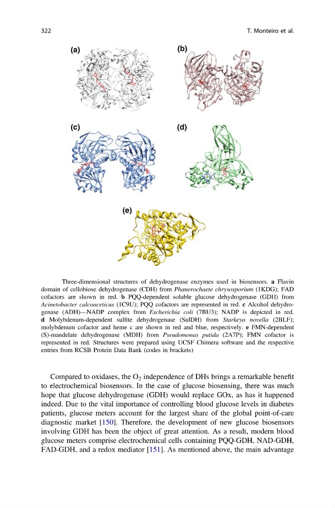
Fig. 6 Three-dimensional structures of dehydrogenase enzymes used in biosensors. a Flavin
domain of cellobiose dehydrogenase (CDH) from Phanerochaete chrysosporium (1KDG); FAD
cofactors are shown in red. b PQQ-dependent soluble glucose dehydrogenase (GDH) from
Acinetobacter calcoaceticus (1C9U); PQQ cofactors are represented in red. c Alcohol dehydrogenase (ADH)—NADP complex from Escherichia coli (7BU3); NADP is depicted in red.
d Molybdenum-dependent sulfite dehydrogenase (SulDH) from Starkeya novella (2BLF);
molybdenum cofactor and heme c are shown in red and blue, respectively. e FMN-dependent
(S)-mandelate dehydrogenase (MDH) from Pseudomonas putida (2A7P); FMN cofactor is
represented in red. Structures were prepared using UCSF Chimera software and the respective
entries from RCSB Protein Data Bank (codes in brackets)
322
T. Monteiro et al.
to electrochemical biosensors. In the case of glucose biosensing, there was much
hope that glucose dehydrogenase (GDH) would replace GOx, as has it happened
indeed. Due to the vital importance of controlling blood glucose levels in diabetes
patients, glucose meters account for the largest share of the global point-of-care
diagnostic market [150]. Therefore, the development of new glucose biosensors
involving GDH has been the object of great attention. As a result, modern blood
glucose meters comprise electrochemical cells containing PQQ-GDH, NAD-GDH,
FAD-GDH, and a redox mediator [151]. As mentioned above, the main advantage
Fig. 6 Three-dimensional structures of dehydrogenase enzymes used in biosensors. a Flavin
domain of cellobiose dehydrogenase (CDH) from Phanerochaete chrysosporium (1KDG); FAD
cofactors are shown in red. b PQQ-dependent soluble glucose dehydrogenase (GDH) from
Acinetobacter calcoaceticus (1C9U); PQQ cofactors are represented in red. c Alcohol dehydrogenase (ADH)—NADP complex from Escherichia coli (7BU3); NADP is depicted in red.
d Molybdenum-dependent sulfite dehydrogenase (SulDH) from Starkeya novella (2BLF);
molybdenum cofactor and heme c are shown in red and blue, respectively. e FMN-dependent
(S)-mandelate dehydrogenase (MDH) from Pseudomonas putida (2A7P); FMN cofactor is
represented in red. Structures were prepared using UCSF Chimera software and the respective
entries from RCSB Protein Data Bank (codes in brackets)
322
T. Monteiro et al.