

3 Active Site Structures and Mechanism of Bimetallic
Hydrogenases
Rarely have the structures of any biomolecular catalyst so obviously indicated
design for their intended function as the hydrogenases. Certainly the entire proteins,
their secondary and tertiary structures, are required for regulating proton and
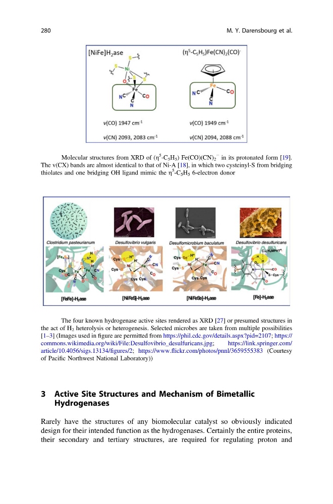
Fig. 4 Molecular structures from XRD of (η
5
-C 5 H 5 ) Fe(CO)(CN) 2
− in its protonated form [19].
The v(CX) bands are almost identical to that of Ni-A [18], in which two cysteinyl-S from bridging
thiolates and one bridging OH ligand mimic the η
5
-C 5 H 5 6-electron donor
Fig. 5 The four known hydrogenase active sites rendered as XRD [27] or presumed structures in
the act of H 2 heterolysis or heterogenesis. Selected microbes are taken from multiple possibilities
[1–3] (Images used in figure are permitted from https://phil.cdc.gov/details.aspx?pid=2107; https://
commons.wikimedia.org/wiki/File:Desulfovibrio_desulfuricans.jpg;
https://link.springer.com/
article/10.4056/sigs.13134/figures/2; https://www.flickr.com/photos/pnnl/3659555383 (Courtesy
of Pacific Northwest National Laboratory))
280
M. Y. Darensbourg et al.
Hydrogenases
Rarely have the structures of any biomolecular catalyst so obviously indicated
design for their intended function as the hydrogenases. Certainly the entire proteins,
their secondary and tertiary structures, are required for regulating proton and
Fig. 4 Molecular structures from XRD of (η
5
-C 5 H 5 ) Fe(CO)(CN) 2
− in its protonated form [19].
The v(CX) bands are almost identical to that of Ni-A [18], in which two cysteinyl-S from bridging
thiolates and one bridging OH ligand mimic the η
5
-C 5 H 5 6-electron donor
Fig. 5 The four known hydrogenase active sites rendered as XRD [27] or presumed structures in
the act of H 2 heterolysis or heterogenesis. Selected microbes are taken from multiple possibilities
[1–3] (Images used in figure are permitted from https://phil.cdc.gov/details.aspx?pid=2107; https://
commons.wikimedia.org/wiki/File:Desulfovibrio_desulfuricans.jpg;
https://link.springer.com/
article/10.4056/sigs.13134/figures/2; https://www.flickr.com/photos/pnnl/3659555383 (Courtesy
of Pacific Northwest National Laboratory))
280
M. Y. Darensbourg et al.