

ppm V
À1 for
13 CN have been observed. The fact that the changes observed under
potential control are entirely due to the Knight shift is confirmed by comparing the
13 C chemical shift response of CO to an electric field in biomolecules, which is of
the order of À15 ppm V
À1
. The changes in 2p
à back donation are accounted for by
the variations in the vibrational frequency, n CO . The CO stretch frequency is lowered when the back donation is high. The above points suggest that the
13 C NMR
shift and the vibrational frequency of the adsorbed CO are electronic in nature.
This originates from the changes in E f –LDOS at the nanocrystalline metal surface
and at the adsorbate, induced by electrochemical potential control [142]. A layer
model analysis is used to describe the
195 Pt NMR spectrum of nanoscopic materials [144]. It is also possible to correlate the electronegativity of the adsorbates with
the Knight shift associated with the Pt nanoparticles [138]. The orientation of adsorbates on metallic substrates under potential control conditions has also been
explored [122, 131]. Tong and co-workers have recently demonstrated the use of
EC-NMR to investigate the electronic environment of the core of MPCs [145].
Other spectroscopic techniques that have been used with electrochemistry to
probe nanoparticles include electronic and vibrational spectroscopies. The spectroelectrochemistry of nanosized silver particles based on their interaction with
planar electrodes has been studied recently [146] using optically transparent thin
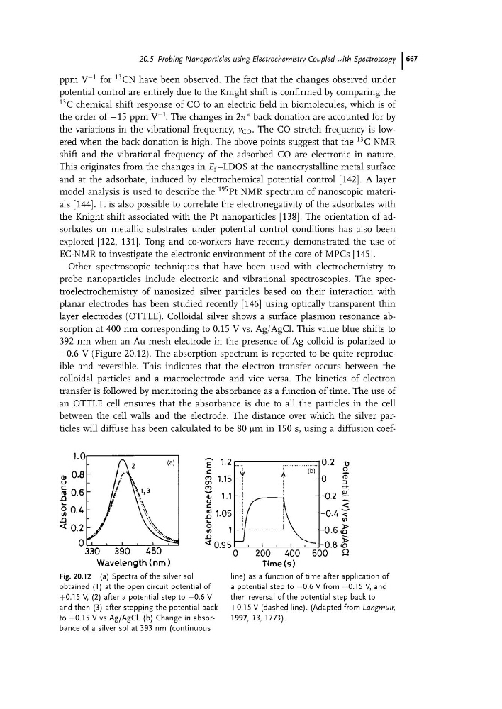
layer electrodes (OTTLE). Colloidal silver shows a surface plasmon resonance absorption at 400 nm corresponding to 0.15 V vs. Ag/AgCl. This value blue shifts to
392 nm when an Au mesh electrode in the presence of Ag colloid is polarized to
À0.6 V (Figure 20.12). The absorption spectrum is reported to be quite reproducible and reversible. This indicates that the electron transfer occurs between the
colloidal particles and a macroelectrode and vice versa. The kinetics of electron
transfer is followed by monitoring the absorbance as a function of time. The use of
an OTTLE cell ensures that the absorbance is due to all the particles in the cell
between the cell walls and the electrode. The distance over which the silver particles will diffuse has been calculated to be 80 mm in 150 s, using a diffusion coefFig. 20.12 (a) Spectra of the silver sol
obtained (1) at the open circuit potential of
þ0.15 V, (2) after a potential step to À0.6 V
and then (3) after stepping the potential back
to þ0.15 V vs Ag/AgCl. (b) Change in absorbance of a silver sol at 393 nm (continuous
line) as a function of time after application of
a potential step to À0.6 V from þ0.15 V, and
then reversal of the potential step back to
þ0.15 V (dashed line). (Adapted from Langmuir,
1997, 13, 1773).
20.5 Probing Nanoparticles using Electrochemistry Coupled with Spectroscopy 667