

monitoring applications is enhanced by the inert surface coating, which renders
QDs less toxic than organic dyes.
Another key advantage is the use of multicolor nanocrystals to simultaneously
image several targets inside living cells or on the cell surface [49]. This is especially
useful in the early detection and identification of cancer tumors based on multiple
markers. An extension of this type of application is the use of near-infrared (NIR)
QDs for in vivo molecular imaging. Similar long-wavelength dyes based on organic
NIR fluorophores have been used for tumor imaging in live animals [51, 52]. Recent research has demonstrated that NIR fluorescent nanocrystals having emission
wavelengths as long as 850 nm and quantum yields as high as 50% at room temperature can be synthesized and they are compatible with the bioconjugation
techniques developed for (CdSe)ZnS QDs. In comparison with carbocyanine and
related organic dyes, these NIR QDs are much brighter (higher quantum yields
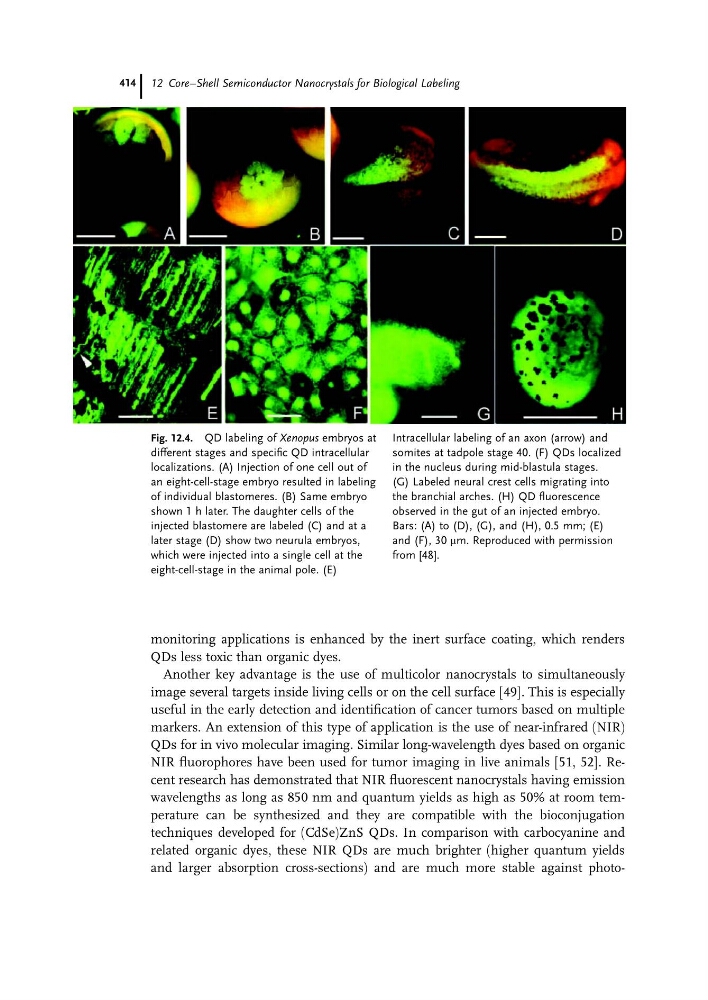
and larger absorption cross-sections) and are much more stable against photoFig. 12.4. QD labeling of Xenopus embryos at
different stages and specific QD intracellular
localizations. (A) Injection of one cell out of
an eight-cell-stage embryo resulted in labeling
of individual blastomeres. (B) Same embryo
shown 1 h later. The daughter cells of the
injected blastomere are labeled (C) and at a
later stage (D) show two neurula embryos,
which were injected into a single cell at the
eight-cell-stage in the animal pole. (E)
Intracellular labeling of an axon (arrow) and
somites at tadpole stage 40. (F) QDs localized
in the nucleus during mid-blastula stages.
(G) Labeled neural crest cells migrating into
the branchial arches. (H) QD fluorescence
observed in the gut of an injected embryo.
Bars: (A) to (D), (G), and (H), 0.5 mm; (E)
and (F), 30 mm. Reproduced with permission
from [48].
12 Core–Shell Semiconductor Nanocrystals for Biological Labeling
414
QDs less toxic than organic dyes.
Another key advantage is the use of multicolor nanocrystals to simultaneously
image several targets inside living cells or on the cell surface [49]. This is especially
useful in the early detection and identification of cancer tumors based on multiple
markers. An extension of this type of application is the use of near-infrared (NIR)
QDs for in vivo molecular imaging. Similar long-wavelength dyes based on organic
NIR fluorophores have been used for tumor imaging in live animals [51, 52]. Recent research has demonstrated that NIR fluorescent nanocrystals having emission
wavelengths as long as 850 nm and quantum yields as high as 50% at room temperature can be synthesized and they are compatible with the bioconjugation
techniques developed for (CdSe)ZnS QDs. In comparison with carbocyanine and
related organic dyes, these NIR QDs are much brighter (higher quantum yields
and larger absorption cross-sections) and are much more stable against photoFig. 12.4. QD labeling of Xenopus embryos at
different stages and specific QD intracellular
localizations. (A) Injection of one cell out of
an eight-cell-stage embryo resulted in labeling
of individual blastomeres. (B) Same embryo
shown 1 h later. The daughter cells of the
injected blastomere are labeled (C) and at a
later stage (D) show two neurula embryos,
which were injected into a single cell at the
eight-cell-stage in the animal pole. (E)
Intracellular labeling of an axon (arrow) and
somites at tadpole stage 40. (F) QDs localized
in the nucleus during mid-blastula stages.
(G) Labeled neural crest cells migrating into
the branchial arches. (H) QD fluorescence
observed in the gut of an injected embryo.
Bars: (A) to (D), (G), and (H), 0.5 mm; (E)
and (F), 30 mm. Reproduced with permission
from [48].
12 Core–Shell Semiconductor Nanocrystals for Biological Labeling
414