

6. Prior to addition of samples, wash the plates 5Â with 200 μL
per well of Wash Buffer solution using the automated plate
washer.
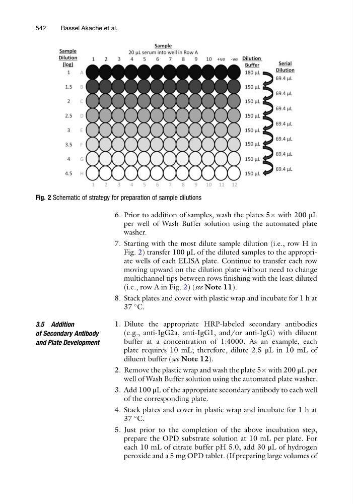
7. Starting with the most dilute sample dilution (i.e., row H in
Fig. 2) transfer 100 μL of the diluted samples to the appropriate wells of each ELISA plate. Continue to transfer each row
moving upward on the dilution plate without need to change
multichannel tips between rows finishing with the least diluted
(i.e., row A in Fig. 2) (see Note 11).
8. Stack plates and cover with plastic wrap and incubate for 1 h at
37
C.
3.5 Addition
of Secondary Antibody
and Plate Development
1. Dilute the appropriate HRP-labeled secondary antibodies
(e.g., anti-IgG2a, anti-IgG1, and/or anti-IgG) with diluent
buffer at a concentration of 1:4000. As an example, each
plate requires 10 mL; therefore, dilute 2.5 μL in 10 mL of
diluent buffer (see Note 12).
2. Remove the plastic wrap and wash the plate 5Â with 200 μL per
well of Wash Buffer solution using the automated plate washer.
3. Add 100 μL of the appropriate secondary antibody to each well
of the corresponding plate.
4. Stack plates and cover in plastic wrap and incubate for 1 h at
37
C.
5. Just prior to the completion of the above incubation step,
prepare the OPD substrate solution at 10 mL per plate. For
each 10 mL of citrate buffer pH 5.0, add 30 μL of hydrogen
peroxide and a 5 mg OPD tablet. (If preparing large volumes of
Fig. 2 Schematic of strategy for preparation of sample dilutions
542
Bassel Akache et al.
per well of Wash Buffer solution using the automated plate
washer.
7. Starting with the most dilute sample dilution (i.e., row H in
Fig. 2) transfer 100 μL of the diluted samples to the appropriate wells of each ELISA plate. Continue to transfer each row
moving upward on the dilution plate without need to change
multichannel tips between rows finishing with the least diluted
(i.e., row A in Fig. 2) (see Note 11).
8. Stack plates and cover with plastic wrap and incubate for 1 h at
37
C.
3.5 Addition
of Secondary Antibody
and Plate Development
1. Dilute the appropriate HRP-labeled secondary antibodies
(e.g., anti-IgG2a, anti-IgG1, and/or anti-IgG) with diluent
buffer at a concentration of 1:4000. As an example, each
plate requires 10 mL; therefore, dilute 2.5 μL in 10 mL of
diluent buffer (see Note 12).
2. Remove the plastic wrap and wash the plate 5Â with 200 μL per
well of Wash Buffer solution using the automated plate washer.
3. Add 100 μL of the appropriate secondary antibody to each well
of the corresponding plate.
4. Stack plates and cover in plastic wrap and incubate for 1 h at
37
C.
5. Just prior to the completion of the above incubation step,
prepare the OPD substrate solution at 10 mL per plate. For
each 10 mL of citrate buffer pH 5.0, add 30 μL of hydrogen
peroxide and a 5 mg OPD tablet. (If preparing large volumes of
Fig. 2 Schematic of strategy for preparation of sample dilutions
542
Bassel Akache et al.