

2.4 Lymph Node
Collection
and Processing
1. R10 complete medium.
2. Sterilized frosted microscope slides.
3. Sterilized petri dishes.
4. Microcentrifuge tubes, 1.5 mL.
2.5 Cell Counting
1. Cellometer Auto 2000 cell viability counter (Nexcelom
Bioscience).
2. Cellometer disposable counting chambers (Nexcelom
Bioscience).
3. ViaStain™ Acridine Orange/Propidium Iodide (AO/PI) viability stain (Nexcelom Bioscience).
2.6 Flow Cytometry
1. Viability Dye: LIVE/DEAD™ Fixable Dead Cell Stain kit
(Invitrogen).
2. Fluorochrome-conjugated antibodies: anti-mouse CD45
(30-F11), CD11b (M1/70), Ly-6G (1A8), Ly-6C (HK1.4),
F4/80 (BM8), CD11c (HL3), CD45R/B220 (RA3-6B2),
CD8a (53-6.7) (see Note 4).
3. UltraComp eBeads™ compensation beads (Invitrogen).
4. Staining buffer: 2% FBS and 3 mM EDTA in 1Â PBS.
5. Fc block: 20 μg anti-mouse CD16/32 in 1 mL staining buffer.
6. Cytofix™ fixation buffer (BD Biosciences).
7. Round-bottom polystyrene tubes, 5 mL.
8. 96-Well DeepWell™ polypropylene microplates, 2 mL.
9. Flow Cytometer: LSRFortessa™ (BD Biosciences).
3 Methods
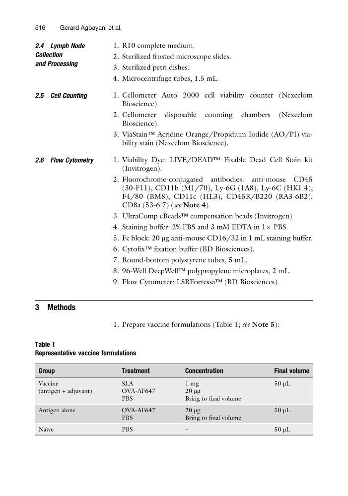
1. Prepare vaccine formulations (Table 1; see Note 5):
Table 1
Representative vaccine formulations
Group
Treatment
Concentration
Final volume
Vaccine
(antigen + adjuvant)
SLA
1 mg
50 μL
OVA-AF647
20 μg
PBS
Bring to final volume
Antigen alone
OVA-AF647
20 μg
5 0 μL
PBS
Bring to final volume
Naı ¨ve
PBS
–
50 μL
516
Gerard Agbayani et al.
Collection
and Processing
1. R10 complete medium.
2. Sterilized frosted microscope slides.
3. Sterilized petri dishes.
4. Microcentrifuge tubes, 1.5 mL.
2.5 Cell Counting
1. Cellometer Auto 2000 cell viability counter (Nexcelom
Bioscience).
2. Cellometer disposable counting chambers (Nexcelom
Bioscience).
3. ViaStain™ Acridine Orange/Propidium Iodide (AO/PI) viability stain (Nexcelom Bioscience).
2.6 Flow Cytometry
1. Viability Dye: LIVE/DEAD™ Fixable Dead Cell Stain kit
(Invitrogen).
2. Fluorochrome-conjugated antibodies: anti-mouse CD45
(30-F11), CD11b (M1/70), Ly-6G (1A8), Ly-6C (HK1.4),
F4/80 (BM8), CD11c (HL3), CD45R/B220 (RA3-6B2),
CD8a (53-6.7) (see Note 4).
3. UltraComp eBeads™ compensation beads (Invitrogen).
4. Staining buffer: 2% FBS and 3 mM EDTA in 1Â PBS.
5. Fc block: 20 μg anti-mouse CD16/32 in 1 mL staining buffer.
6. Cytofix™ fixation buffer (BD Biosciences).
7. Round-bottom polystyrene tubes, 5 mL.
8. 96-Well DeepWell™ polypropylene microplates, 2 mL.
9. Flow Cytometer: LSRFortessa™ (BD Biosciences).
3 Methods
1. Prepare vaccine formulations (Table 1; see Note 5):
Table 1
Representative vaccine formulations
Group
Treatment
Concentration
Final volume
Vaccine
(antigen + adjuvant)
SLA
1 mg
50 μL
OVA-AF647
20 μg
PBS
Bring to final volume
Antigen alone
OVA-AF647
20 μg
5 0 μL
PBS
Bring to final volume
Naı ¨ve
PBS
–
50 μL
516
Gerard Agbayani et al.