

2. 0.2% Collagenase Type IV digestion buffer: Dissolve 0.2 g in
100 mL 1Â Hank’s Balanced Salt Solution (HBSS) containing
2% FBS. Filter-sterilize using 0.2 μm membrane filter. Store at
À20
C. Keep on ice or at 4
C upon thawing before use.
3. Sterilized surgical scissors.
4. Sterilized surgical forceps.
5. Sterilized petri dishes.
6. Microcentrifuge tubes, 1.5 mL.
7. Falcon™ conical centrifuge tubes, 15 mL.
8. Falcon™ conical centrifuge tubes, 50 mL.
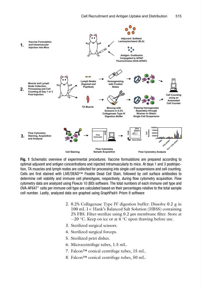
Fig. 1 Schematic overview of experimental procedures. Vaccine formulations are prepared according to
optimal adjuvant and antigen concentrations and injected intramuscularly to mice. At days 1 and 3 postinjection, TA muscles and lymph nodes are collected for processing into single-cell suspensions and cell counting.
Cells are first stained with LIVE/DEAD™ Fixable Dead Cell Stain, followed by cell surface antibodies to
determine cell viability and immune cell phenotypes, respectively, during flow cytometry acquisition. Flow
cytometry data are analyzed using FlowJo 10 (BD) software. The total numbers of each immune cell type and
OVA-AF647
+ cells per immune cell type are calculated based on their percentages relative to the total sample
cell number. Lastly, analyzed data are graphed using GraphPad® Prism 8 software
Cell Recruitment and Antigen Uptake and Distribution
515
100 mL 1Â Hank’s Balanced Salt Solution (HBSS) containing
2% FBS. Filter-sterilize using 0.2 μm membrane filter. Store at
À20
C. Keep on ice or at 4
C upon thawing before use.
3. Sterilized surgical scissors.
4. Sterilized surgical forceps.
5. Sterilized petri dishes.
6. Microcentrifuge tubes, 1.5 mL.
7. Falcon™ conical centrifuge tubes, 15 mL.
8. Falcon™ conical centrifuge tubes, 50 mL.
Fig. 1 Schematic overview of experimental procedures. Vaccine formulations are prepared according to
optimal adjuvant and antigen concentrations and injected intramuscularly to mice. At days 1 and 3 postinjection, TA muscles and lymph nodes are collected for processing into single-cell suspensions and cell counting.
Cells are first stained with LIVE/DEAD™ Fixable Dead Cell Stain, followed by cell surface antibodies to
determine cell viability and immune cell phenotypes, respectively, during flow cytometry acquisition. Flow
cytometry data are analyzed using FlowJo 10 (BD) software. The total numbers of each immune cell type and
OVA-AF647
+ cells per immune cell type are calculated based on their percentages relative to the total sample
cell number. Lastly, analyzed data are graphed using GraphPad® Prism 8 software
Cell Recruitment and Antigen Uptake and Distribution
515