

(e) Set up data collection parameters and conditions in data
collection software (Recorder). The low dose imaging
mode should be applied, in which the position for search
and focus is set in nonacquisition region.
(f) Perform the calibrations for beam brightness, compensators (beam shift/tilt balance and image shift), and stage
condition etc. at recording magnification at 0
.
(g) Find a typical effective region where the antigen capsulated archaeosomes is in the field of view and also the
tilting angle is in the range of Æ70
(Fig. 6).
(h) Tilt specimen holder in X-axis to the maximum minus
angle and start the image acquisition automatically till
the tilt angle reaching the maximum plus.
Fig. 6 Cryo-TEM bright field image of archaeosomes entrapped with OVA
immunolabeled with 1.4 nm gold conjugated Ab. The archaeosomes in the
field of view were used for tilt-series micrograph
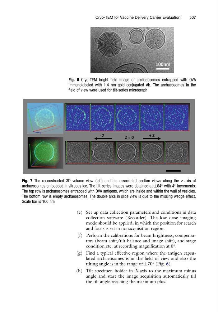
Fig. 7 The reconstructed 3D volume view (left) and the associated section views along the z axis of
archaeosomes embedded in vitreous ice. The tilt-series images were obtained at Æ64
with 4
increments.
The top row is archaeosomes entrapped with OVA antigens, which are inside and within the wall of vesicles.
The bottom row is empty archaeosomes. The double arcs in slice view is due to the missing wedge effect.
Scale bar is 100 nm
Cryo-TEM for Vaccine Delivery Carrier Evaluation
507
collection software (Recorder). The low dose imaging
mode should be applied, in which the position for search
and focus is set in nonacquisition region.
(f) Perform the calibrations for beam brightness, compensators (beam shift/tilt balance and image shift), and stage
condition etc. at recording magnification at 0
.
(g) Find a typical effective region where the antigen capsulated archaeosomes is in the field of view and also the
tilting angle is in the range of Æ70
(Fig. 6).
(h) Tilt specimen holder in X-axis to the maximum minus
angle and start the image acquisition automatically till
the tilt angle reaching the maximum plus.
Fig. 6 Cryo-TEM bright field image of archaeosomes entrapped with OVA
immunolabeled with 1.4 nm gold conjugated Ab. The archaeosomes in the
field of view were used for tilt-series micrograph
Fig. 7 The reconstructed 3D volume view (left) and the associated section views along the z axis of
archaeosomes embedded in vitreous ice. The tilt-series images were obtained at Æ64
with 4
increments.
The top row is archaeosomes entrapped with OVA antigens, which are inside and within the wall of vesicles.
The bottom row is empty archaeosomes. The double arcs in slice view is due to the missing wedge effect.
Scale bar is 100 nm
Cryo-TEM for Vaccine Delivery Carrier Evaluation
507