

(b) Choose the proper magnification for imaging the archaeosomes (see Note 12).
(c) Adjust the electron emitter parameters and measure the
beam current if necessary.
(d) Adjust and optimize the image contrast by inserting
objective aperture and a 10 eV energy filter slit.
(e) Adjust the illumination area on the specimen. Optimize
parameters in the setting for Gatan slow-scan 2k  2k
camera including the binning of 2 Â 2 or 4 Â 4 in search
mode and 1 Â 1 binning in acquisition mode. Set acquisition time to obtain the archaeosomes image with beam
dose of 2 e
À
/A ˚ 2 .
(f) Focus the specimen and blank the beam.
(g) Move the stage to the new area nearby (not previously
exposed in the beam) and wait for several seconds to
1 min.
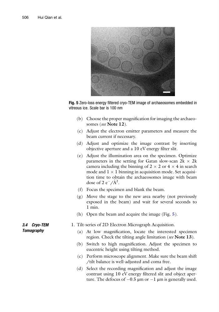
(h) Open the beam and acquire the image (Fig. 5).
3.4 Cryo-TEM
Tomography
1. Tilt-series of 2D Electron Micrograph Acquisition.
(a) At low magnification, locate the interested specimen
region. Check the tilting angle limitation (see Note 13).
(b) Switch to high magnification. Adjust the specimen to
eucentric height using tilting method.
(c) Perform microscope alignment. Make sure the beam shift
/tilt balance is well-adjusted and coma free.
(d) Select the recording magnification and adjust the image
contrast using 10 eV energy filtered slit and object aperture. The defocus of À0.5 μm or À1 μm is generally used.
Fig. 5 Zero-loss energy filtered cryo-TEM image of archaeosomes embedded in
vitreous ice. Scale bar is 100 nm
506
Hui Qian et al.
(c) Adjust the electron emitter parameters and measure the
beam current if necessary.
(d) Adjust and optimize the image contrast by inserting
objective aperture and a 10 eV energy filter slit.
(e) Adjust the illumination area on the specimen. Optimize
parameters in the setting for Gatan slow-scan 2k  2k
camera including the binning of 2 Â 2 or 4 Â 4 in search
mode and 1 Â 1 binning in acquisition mode. Set acquisition time to obtain the archaeosomes image with beam
dose of 2 e
À
/A ˚ 2 .
(f) Focus the specimen and blank the beam.
(g) Move the stage to the new area nearby (not previously
exposed in the beam) and wait for several seconds to
1 min.
(h) Open the beam and acquire the image (Fig. 5).
3.4 Cryo-TEM
Tomography
1. Tilt-series of 2D Electron Micrograph Acquisition.
(a) At low magnification, locate the interested specimen
region. Check the tilting angle limitation (see Note 13).
(b) Switch to high magnification. Adjust the specimen to
eucentric height using tilting method.
(c) Perform microscope alignment. Make sure the beam shift
/tilt balance is well-adjusted and coma free.
(d) Select the recording magnification and adjust the image
contrast using 10 eV energy filtered slit and object aperture. The defocus of À0.5 μm or À1 μm is generally used.
Fig. 5 Zero-loss energy filtered cryo-TEM image of archaeosomes embedded in
vitreous ice. Scale bar is 100 nm
506
Hui Qian et al.