

Here, using archaeosomes as a model lipid-base nanoparticle
and ovalbumin (OVA) as a model protein antigen, we describe the
plunge freezing method for archaeosome-based formulations in
aqueous solution and combine it with immunogold labeling techniques to identify OVA antigens in the formulation. Cryo-TEM
bright field imaging and electron tomography using the single tiltseries method reveals the archaeosome morphology and the antigen distribution in 3D [7]. The techniques described herein can
also be applied to other nanocarriers and antigens.
2 Materials
2.1 Vaccine Delivery
Carriers
1. Sulfated lactosylarchaeol (SLA; 6
0 -sulfate-β-D-Galp-(1,4)-β-DGlcp-(1,1)-archaeol) glycolipids were prepared in house [8].
2. Ovalbumin antigen (OVA): Type VI; Sigma Aldrich.
3. Ultrasonic water bath; Fisher Scientific, Nepean, ON, Canada.
2.2 Immunogold
Labeling Reagents
1. Antigen Conjugates: polyclonal mouse anti-OVA IgG antibody
is prepared in-house as primary antibody; Nanogold
® -IgG,
1.4 nm (Nanogold conjugated secondary antibody goat antimouse IgG) is purchased from Nanoprobes Inc., Yaphank,
NY, USA.
2. PBS-BSA buffer: 20 mM phosphate, 150 mM NaCl, pH 7.4,
0.5% BSA, 0.1% gelatin.
3. PBS buffer: 20 mM phosphate, 150 mM NaCl, pH 7.4.
2.3 TEM Specimen
Preparation Materials
1. TEM grids: ultrathin carbon film (3–4 nm thick) supported
TEM grids (400 mesh), lacey carbon film with an ultrathin
formvar film (3–4 nm) Nickle (400 mesh), lacey carbon copper
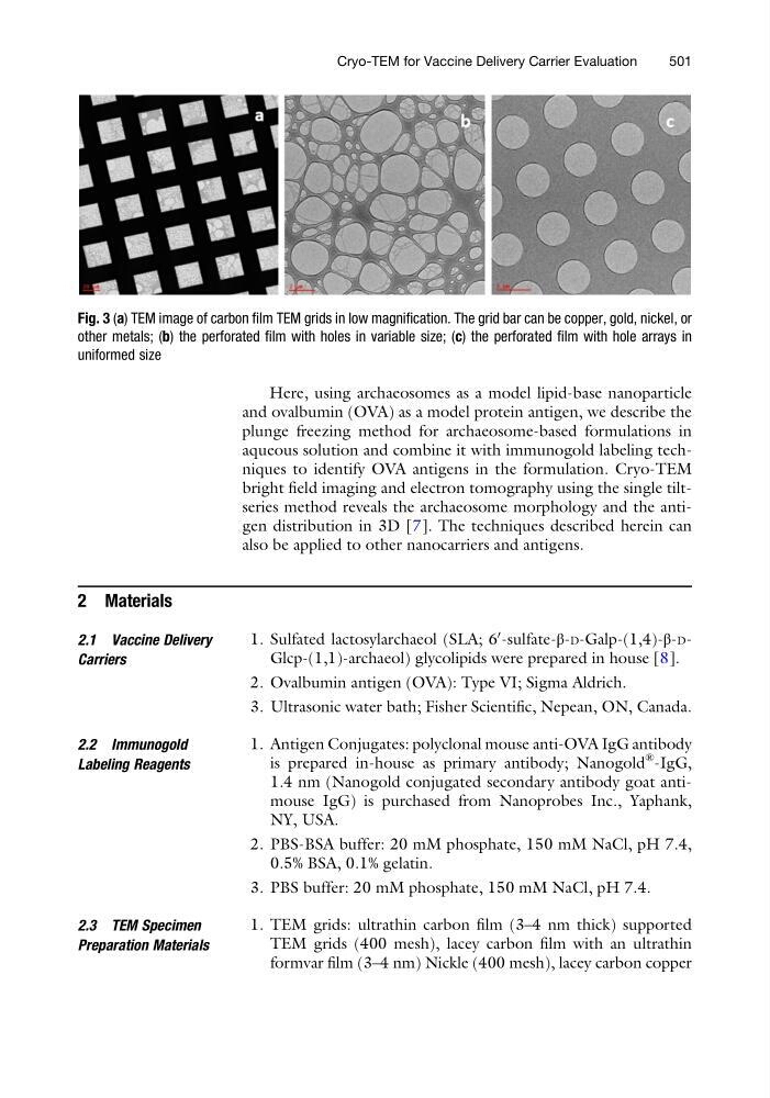
Fig. 3 (a) TEM image of carbon film TEM grids in low magnification. The grid bar can be copper, gold, nickel, or
other metals; (b) the perforated film with holes in variable size; (c) the perforated film with hole arrays in
uniformed size
Cryo-TEM for Vaccine Delivery Carrier Evaluation
501
and ovalbumin (OVA) as a model protein antigen, we describe the
plunge freezing method for archaeosome-based formulations in
aqueous solution and combine it with immunogold labeling techniques to identify OVA antigens in the formulation. Cryo-TEM
bright field imaging and electron tomography using the single tiltseries method reveals the archaeosome morphology and the antigen distribution in 3D [7]. The techniques described herein can
also be applied to other nanocarriers and antigens.
2 Materials
2.1 Vaccine Delivery
Carriers
1. Sulfated lactosylarchaeol (SLA; 6
0 -sulfate-β-D-Galp-(1,4)-β-DGlcp-(1,1)-archaeol) glycolipids were prepared in house [8].
2. Ovalbumin antigen (OVA): Type VI; Sigma Aldrich.
3. Ultrasonic water bath; Fisher Scientific, Nepean, ON, Canada.
2.2 Immunogold
Labeling Reagents
1. Antigen Conjugates: polyclonal mouse anti-OVA IgG antibody
is prepared in-house as primary antibody; Nanogold
® -IgG,
1.4 nm (Nanogold conjugated secondary antibody goat antimouse IgG) is purchased from Nanoprobes Inc., Yaphank,
NY, USA.
2. PBS-BSA buffer: 20 mM phosphate, 150 mM NaCl, pH 7.4,
0.5% BSA, 0.1% gelatin.
3. PBS buffer: 20 mM phosphate, 150 mM NaCl, pH 7.4.
2.3 TEM Specimen
Preparation Materials
1. TEM grids: ultrathin carbon film (3–4 nm thick) supported
TEM grids (400 mesh), lacey carbon film with an ultrathin
formvar film (3–4 nm) Nickle (400 mesh), lacey carbon copper
Fig. 3 (a) TEM image of carbon film TEM grids in low magnification. The grid bar can be copper, gold, nickel, or
other metals; (b) the perforated film with holes in variable size; (c) the perforated film with hole arrays in
uniformed size
Cryo-TEM for Vaccine Delivery Carrier Evaluation
501