

3. Compare the fluorescence measurements to a GFP standard
curve and determine the fraction bound by comparing to initial
amount of introduced protein: Binding efficiency ¼ (F sample À F blank )/(F max À F blank ) * 100%.
3.4 Polysaccharide
Encapsulation
Efficiency
1. Add 0.6 mL of fully assembled LEPS construct to a glass vial.
In addition, a negative control containing a LEPS construct
without polysaccharides should be included. Furthermore, a
standard curve of polysaccharides ranging from 0 to 0.6 mg/
mL should be included.
2. Next, add 0.3 mL of phenol 5% (w/v) to the vial.
3. Add 1.5 mL concentrated sulfuric acid to the mixture.
4. After adding each component, mix the solution by vortexing
for 5 s.
5. Following mixing, transfer 250 μL of sample to a microplate.
6. Seal the microplate and then place it into a sealable plastic bag.
7. Incubate the microplate in a 92
C water bath for 15 min (see
Note 4).
8. Transfer the microplate to a room temperature water bath
(20–25
C) and incubate for an additional 15 min.
9. Measure the absorbance at 480 nm using a plate reader (see
Note 5). Utilize a standard curve to determine the concentration of polysaccharide in each well.
10. Compare the measured values to that obtained for the initial
concentration of introduced polysaccharide (0.6 mg, A max )
and the blank containing no polysaccharides (A blank ) to determine encapsulation efficiency using the following calculation:
Encapsulation
efficiency
¼
(A sample
À
A blank )/
(A max À A blank ) * 100%.
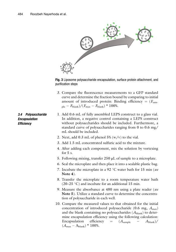
Fig. 3 Liposome polysaccharide encapsulation, surface protein attachment, and
purification steps
484
Roozbeh Nayerhoda et al.
curve and determine the fraction bound by comparing to initial
amount of introduced protein: Binding efficiency ¼ (F sample À F blank )/(F max À F blank ) * 100%.
3.4 Polysaccharide
Encapsulation
Efficiency
1. Add 0.6 mL of fully assembled LEPS construct to a glass vial.
In addition, a negative control containing a LEPS construct
without polysaccharides should be included. Furthermore, a
standard curve of polysaccharides ranging from 0 to 0.6 mg/
mL should be included.
2. Next, add 0.3 mL of phenol 5% (w/v) to the vial.
3. Add 1.5 mL concentrated sulfuric acid to the mixture.
4. After adding each component, mix the solution by vortexing
for 5 s.
5. Following mixing, transfer 250 μL of sample to a microplate.
6. Seal the microplate and then place it into a sealable plastic bag.
7. Incubate the microplate in a 92
C water bath for 15 min (see
Note 4).
8. Transfer the microplate to a room temperature water bath
(20–25
C) and incubate for an additional 15 min.
9. Measure the absorbance at 480 nm using a plate reader (see
Note 5). Utilize a standard curve to determine the concentration of polysaccharide in each well.
10. Compare the measured values to that obtained for the initial
concentration of introduced polysaccharide (0.6 mg, A max )
and the blank containing no polysaccharides (A blank ) to determine encapsulation efficiency using the following calculation:
Encapsulation
efficiency
¼
(A sample
À
A blank )/
(A max À A blank ) * 100%.
Fig. 3 Liposome polysaccharide encapsulation, surface protein attachment, and
purification steps
484
Roozbeh Nayerhoda et al.