

7. Resuspend the purified liposome in up to 500 μL PBS and
transfer the solution to a 1.5 mL centrifugal tube.
8. Add 140 μg of GFP to the liposome solution (see Note 3) and
incubate for 30 min at room temperature (20–25
C).
9. Transfer 500 μL of the sample to the 300 kDa microcentrifuge
tubes and centrifuge for 5 min at 4
C at 12,000 rcf.
10. Resuspend the final purified liposomes in up to 500 μL of PBS
and transfer to a microcentrifuge tube and repeat the centrifugation for 5 min at 4
C at 12,000 rcf.
11. Resuspend the purified liposome in up to 500 μL PBS and
transfer the solution to a 1.5 mL centrifugal tube (Fig. 3).
3.3 Measuring
Protein Binding
Efficiency
1. Transfer 50 μL of liposome-bound GFP to a 96-well plate. A
negative control of the liposome without added GFP should be
included. In addition, a positive control containing the amount
of GFP initially added should be included.
2. Measure the fluorescence of the plate wells using a plate reader
with at an excitation wavelength of 359 nm and an emission
wavelength of 508 nm. The fluorescence of the liposome without added GFP (F blank ) and the fluorescence of the amount of
GFP initially to the liposomes added F max should be noted.
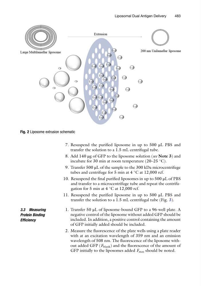
Fig. 2 Liposome extrusion schematic
Liposomal Dual Antigen Delivery
483
transfer the solution to a 1.5 mL centrifugal tube.
8. Add 140 μg of GFP to the liposome solution (see Note 3) and
incubate for 30 min at room temperature (20–25
C).
9. Transfer 500 μL of the sample to the 300 kDa microcentrifuge
tubes and centrifuge for 5 min at 4
C at 12,000 rcf.
10. Resuspend the final purified liposomes in up to 500 μL of PBS
and transfer to a microcentrifuge tube and repeat the centrifugation for 5 min at 4
C at 12,000 rcf.
11. Resuspend the purified liposome in up to 500 μL PBS and
transfer the solution to a 1.5 mL centrifugal tube (Fig. 3).
3.3 Measuring
Protein Binding
Efficiency
1. Transfer 50 μL of liposome-bound GFP to a 96-well plate. A
negative control of the liposome without added GFP should be
included. In addition, a positive control containing the amount
of GFP initially added should be included.
2. Measure the fluorescence of the plate wells using a plate reader
with at an excitation wavelength of 359 nm and an emission
wavelength of 508 nm. The fluorescence of the liposome without added GFP (F blank ) and the fluorescence of the amount of
GFP initially to the liposomes added F max should be noted.
Fig. 2 Liposome extrusion schematic
Liposomal Dual Antigen Delivery
483