

the volume needed for dilutions. Usually, a dilution of 1:5 is
needed.
l
Prepare standards, as specified by the manufacturer (see Note
14). Prepare enough volume for every new 96-well plate. Standards can be stored at 2–6
C for several weeks.
l
Now, pipet the probes/standard, if necessary, TE buffer for
dilution, and last, diluted Quant-iT™ PicoGreen™ in each
well. Mix the plate thoroughly on the plate shaker for 30 s.
Incubate for 2–5 min, excluded from light. Start reading with
plate reader according to the manufacturer’s instructions (see
Note 15).
Total Protein
l
Prepare standards as described in the manual with BGG or BSA.
Standards can be stored at 2–6
C for several weeks.
l
Prepare working reagent (WR) following the manufacturer’s
instructions for the 96-well plate procedure.
l
Pipet probes/standards and add PBS for a dilution into the wells
containing the probes. A dilution of 1:10 is necessary for probes
containing high loads of protein, for example the supernatant of
lysed cells, or medium-containing serum.
l
Add WR.
l
Now, mix the plate thoroughly on the plate shaker for 30 s.
Incubate for 30 min at 37
C. After letting the plate cool down
to RT, start the reading with the plate reader according to the
manufacturer’s instructions.
Mass Balance Study
l
Determine the vDNA content via qPCR and infectious titer of
the baculovirus via TCID 50 or a plaque assay of the feed, permeate for depth filtration, and retentate and permeate for TFF.
l
By considering the volume/mass of individual fractions, the
vDNA content of the feed is set to be 100%, and fractions should
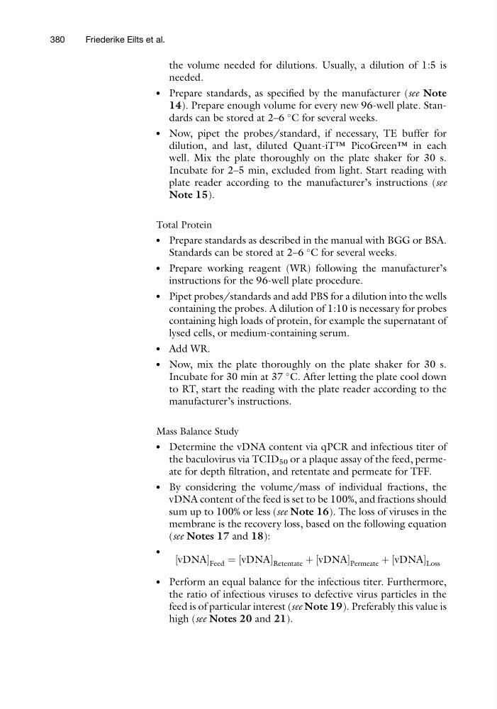
sum up to 100% or less (see Note 16). The loss of viruses in the
membrane is the recovery loss, based on the following equation
(see Notes 17 and 18):
l
vDNA
½
Feed ¼ vDNA
½
Retentate þ vDNA
½
Permeate þ vDNA
½
Loss
l
Perform an equal balance for the infectious titer. Furthermore,
the ratio of infectious viruses to defective virus particles in the
feed is of particular interest (see Note 19). Preferably this value is
high (see Notes 20 and 21).
380
Friederike Eilts et al.
needed.
l
Prepare standards, as specified by the manufacturer (see Note
14). Prepare enough volume for every new 96-well plate. Standards can be stored at 2–6
C for several weeks.
l
Now, pipet the probes/standard, if necessary, TE buffer for
dilution, and last, diluted Quant-iT™ PicoGreen™ in each
well. Mix the plate thoroughly on the plate shaker for 30 s.
Incubate for 2–5 min, excluded from light. Start reading with
plate reader according to the manufacturer’s instructions (see
Note 15).
Total Protein
l
Prepare standards as described in the manual with BGG or BSA.
Standards can be stored at 2–6
C for several weeks.
l
Prepare working reagent (WR) following the manufacturer’s
instructions for the 96-well plate procedure.
l
Pipet probes/standards and add PBS for a dilution into the wells
containing the probes. A dilution of 1:10 is necessary for probes
containing high loads of protein, for example the supernatant of
lysed cells, or medium-containing serum.
l
Add WR.
l
Now, mix the plate thoroughly on the plate shaker for 30 s.
Incubate for 30 min at 37
C. After letting the plate cool down
to RT, start the reading with the plate reader according to the
manufacturer’s instructions.
Mass Balance Study
l
Determine the vDNA content via qPCR and infectious titer of
the baculovirus via TCID 50 or a plaque assay of the feed, permeate for depth filtration, and retentate and permeate for TFF.
l
By considering the volume/mass of individual fractions, the
vDNA content of the feed is set to be 100%, and fractions should
sum up to 100% or less (see Note 16). The loss of viruses in the
membrane is the recovery loss, based on the following equation
(see Notes 17 and 18):
l
vDNA
½
Feed ¼ vDNA
½
Retentate þ vDNA
½
Permeate þ vDNA
½
Loss
l
Perform an equal balance for the infectious titer. Furthermore,
the ratio of infectious viruses to defective virus particles in the
feed is of particular interest (see Note 19). Preferably this value is
high (see Notes 20 and 21).
380
Friederike Eilts et al.