

2. 50 mM HEPES (pH 8.0): Take 50 ml from the stock solution
(100 mM) and increase the volume to 80 ml with DI water.
Adjust the pH to 8.0 using NaOH before increasing the final
volume to 100 ml with DI water. Filter the buffer to sterilize
and store at 4
C.
3. 20 mM HEPES buffer (pH 7.0): Take 20 ml from the stock
solution (100 mM) and increase the volume to 80 ml with DI
water. Adjust the pH to 7.0 using NaOH before increasing the
final volume to 100 ml with DI water. Filter the buffer to
sterilize and store at 4
C.
2.6 Equipment
and Instruments
1. Cell culture incubator (high humidity, 37
C, 5% CO 2 ).
2. Class II biological safety cabinet.
3. Vortex mixer.
4. Water bath at 37
C.
5. Protein Concentrators (PES, 10 kDa MWCO, 0.5 ml).
6. Microcentrifuge.
7. 1000, 200, and 10 μl pipettes.
3 Methods
3.1 Preparing Plates
for Transduction
1. Trypsinize NIH/3T3 cells from a 90% confluent T-25 flask and
resuspend in 8–10 ml of medium.
2. Determine the cell concentration using a hemocytometer and
plate 5 Â 10
4 cells in 500 μl of cell growth medium per well of a
24 well plate.
3. After 18–24 h, the cells should appear 50–60% confluent and
are ready for transduction. (See Table 1 for different seeding
densities and vessel size options).
3.2 Preparing
Adenoviral Stock
1. Decide the desired multiplicity of infection (MOI) for transduction. You will need to know the approximate number of
cells seeded. Refer to Table 1 for seeding densities and volumes
of medium for each well based on the size of the plate (see Note
4).
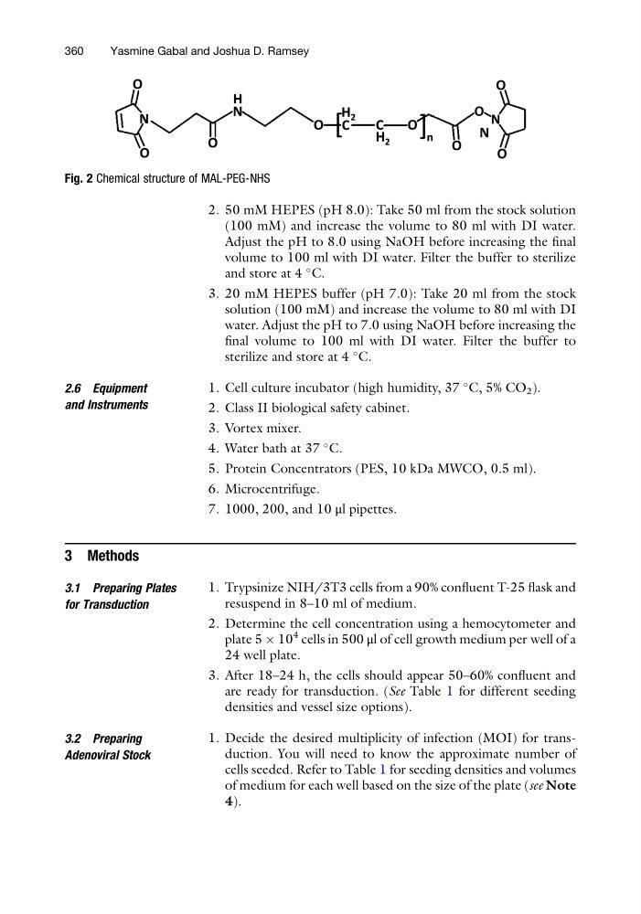
Fig. 2 Chemical structure of MAL-PEG-NHS
360
Yasmine Gabal and Joshua D. Ramsey
(100 mM) and increase the volume to 80 ml with DI water.
Adjust the pH to 8.0 using NaOH before increasing the final
volume to 100 ml with DI water. Filter the buffer to sterilize
and store at 4
C.
3. 20 mM HEPES buffer (pH 7.0): Take 20 ml from the stock
solution (100 mM) and increase the volume to 80 ml with DI
water. Adjust the pH to 7.0 using NaOH before increasing the
final volume to 100 ml with DI water. Filter the buffer to
sterilize and store at 4
C.
2.6 Equipment
and Instruments
1. Cell culture incubator (high humidity, 37
C, 5% CO 2 ).
2. Class II biological safety cabinet.
3. Vortex mixer.
4. Water bath at 37
C.
5. Protein Concentrators (PES, 10 kDa MWCO, 0.5 ml).
6. Microcentrifuge.
7. 1000, 200, and 10 μl pipettes.
3 Methods
3.1 Preparing Plates
for Transduction
1. Trypsinize NIH/3T3 cells from a 90% confluent T-25 flask and
resuspend in 8–10 ml of medium.
2. Determine the cell concentration using a hemocytometer and
plate 5 Â 10
4 cells in 500 μl of cell growth medium per well of a
24 well plate.
3. After 18–24 h, the cells should appear 50–60% confluent and
are ready for transduction. (See Table 1 for different seeding
densities and vessel size options).
3.2 Preparing
Adenoviral Stock
1. Decide the desired multiplicity of infection (MOI) for transduction. You will need to know the approximate number of
cells seeded. Refer to Table 1 for seeding densities and volumes
of medium for each well based on the size of the plate (see Note
4).
Fig. 2 Chemical structure of MAL-PEG-NHS
360
Yasmine Gabal and Joshua D. Ramsey