

pooled sample on the top of the sucrose gradient using a 5 mL
serological pipette. Centrifuge samples at 30,000 rpm
(111,132 Â g, SW41Ti rotor) for 16 h, at 4
C (see Note 16).
6. Collect 750 μL fractions from the top of the gradient and
perform ELISA using the Monolisa HBsAg ULTRA kit.
7. Pool HBV antigen-positive fractions and dialyze 3Â against 3 L
PBS 0.1Â using SnakeSkin Dialysis Tubing with 10 kDa pores
(see Note 17).
8. Freeze-dialyzed samples in liquid N 2 and concentrate by lyophilization (see Note 18).
9. Add sterile water to the protein powder to a final volume of
300 μL/antigen/1700 cm
2 roller bottle.
10. Quantify HBV antigens levels in the concentrated samples by
using Monolisa HBsAg ULTRA kit. Estimate the yields per
gram fresh cell weight.
11. Quantify HBV chimeric antigens by western blot using a standard curve made of known amounts of commercial L protein
(the preS1 antigen, Beacle). A typical example of purified chimeric HBV protein is shown in Fig. 4 (see Note 19).
12. Determine the protein concentration in the concentrated samples by using the BCA protocol and calculate the purity of
HBV antigens (see Note 20).
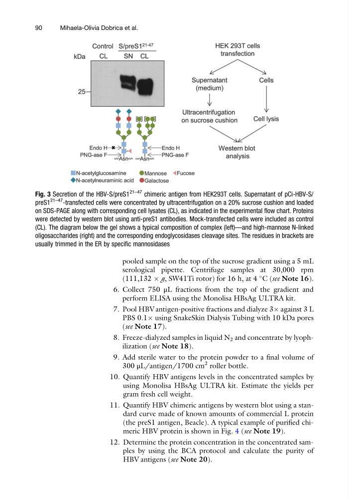
Fig. 3 Secretion of the HBV-S/preS1
21–47 chimeric antigen from HEK293T cells. Supernatant of pCi-HBV-S/
preS1
21–47 -transfected cells were concentrated by ultracentrifugation on a 20% sucrose cushion and loaded
on SDS-PAGE along with corresponding cell lysates (CL), as indicated in the experimental flow chart. Proteins
were detected by western blot using anti-preS1 antibodies. Mock-transfected cells were included as control
(CL). The diagram below the gel shows a typical composition of complex (left)—and high-mannose N-linked
oligosaccharides (right) and the corresponding endoglycosidases cleavage sites. The residues in brackets are
usually trimmed in the ER by specific mannosidases
90
Mihaela-Olivia Dobrica et al.
serological pipette. Centrifuge samples at 30,000 rpm
(111,132 Â g, SW41Ti rotor) for 16 h, at 4
C (see Note 16).
6. Collect 750 μL fractions from the top of the gradient and
perform ELISA using the Monolisa HBsAg ULTRA kit.
7. Pool HBV antigen-positive fractions and dialyze 3Â against 3 L
PBS 0.1Â using SnakeSkin Dialysis Tubing with 10 kDa pores
(see Note 17).
8. Freeze-dialyzed samples in liquid N 2 and concentrate by lyophilization (see Note 18).
9. Add sterile water to the protein powder to a final volume of
300 μL/antigen/1700 cm
2 roller bottle.
10. Quantify HBV antigens levels in the concentrated samples by
using Monolisa HBsAg ULTRA kit. Estimate the yields per
gram fresh cell weight.
11. Quantify HBV chimeric antigens by western blot using a standard curve made of known amounts of commercial L protein
(the preS1 antigen, Beacle). A typical example of purified chimeric HBV protein is shown in Fig. 4 (see Note 19).
12. Determine the protein concentration in the concentrated samples by using the BCA protocol and calculate the purity of
HBV antigens (see Note 20).
Fig. 3 Secretion of the HBV-S/preS1
21–47 chimeric antigen from HEK293T cells. Supernatant of pCi-HBV-S/
preS1
21–47 -transfected cells were concentrated by ultracentrifugation on a 20% sucrose cushion and loaded
on SDS-PAGE along with corresponding cell lysates (CL), as indicated in the experimental flow chart. Proteins
were detected by western blot using anti-preS1 antibodies. Mock-transfected cells were included as control
(CL). The diagram below the gel shows a typical composition of complex (left)—and high-mannose N-linked
oligosaccharides (right) and the corresponding endoglycosidases cleavage sites. The residues in brackets are
usually trimmed in the ER by specific mannosidases
90
Mihaela-Olivia Dobrica et al.