

6. Load samples on SDS-10% PAA gels and electrophorese at
30 mA until the blue dye front reaches the bottom of the gel.
7. Wet nitrocellulose membranes in western-blot transfer buffer
and transfer proteins from gels using a semidry blotter, for
75 min at 60 mA.
8. Block the membranes with 10% nonfat milk in PBS for 1 h.
9. Incubate membranes with mouse anti-preS1 antibody
(1/1000 in antibody dilution buffer), for 1 h (see Note 11).
10. Wash the membranes 3Â with western-blot washing solution,
10 min each time.
11. Incubate membrane with anti-mouse-HRP secondary antibody (1/10,000, in antibody dilution buffer) for 1 h.
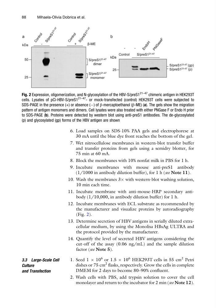
12. Incubate membranes with ECL substrate as recommended by
the manufacturer and visualize proteins by autoradiography
(Fig. 2).
13. Determine secretion of HBV antigens in serially diluted extracellular medium, by using the Monolisa HBsAg ULTRA and
the protocol provided by the manufacturer.
14. Quantify the level of secreted HBV antigens considering the
cut-off of the assay (0.06 ng/mL) and the sample dilution
factor (see Note 5).
3.3 Large-Scale Cell
Culture
and Transfection
1. Seed 1 Â 10
6 or 1.5 Â 10
6 HEK293T cells in 55 cm
2 Petri
dishes or 75 cm
2 flasks, respectively. Grow the cells in complete
DMEM for 2 days to become 80–90% confluent.
2. Wash cells with PBS, add trypsin solution to cover the cell
monolayer and return to the incubator for 2 min (see Note 12).
Fig. 2 Expression, oligomerization, and N-glycosylation of the HBV-S/preS1
21–47 chimeric antigen in HEK293T
cells. Lysates of pCi-HBV-S/preS1
21–47 - or mock-transfected (control) HEK293T cells were subjected to
SDS-PAGE in the presence (+) or absence (À) of β-mercaptoethanol (β-ME) (a). The gels show the migration
pattern of antigen monomers and dimers. Cell lysates were also treated with either PNGase F or Endo H prior
to SDS-PAGE (b). Proteins were detected by western blot using anti-preS1 antibodies. The de-glycosylated
(p) and glycosylated (gp) forms of the HBV antigen are shown
88
Mihaela-Olivia Dobrica et al.
30 mA until the blue dye front reaches the bottom of the gel.
7. Wet nitrocellulose membranes in western-blot transfer buffer
and transfer proteins from gels using a semidry blotter, for
75 min at 60 mA.
8. Block the membranes with 10% nonfat milk in PBS for 1 h.
9. Incubate membranes with mouse anti-preS1 antibody
(1/1000 in antibody dilution buffer), for 1 h (see Note 11).
10. Wash the membranes 3Â with western-blot washing solution,
10 min each time.
11. Incubate membrane with anti-mouse-HRP secondary antibody (1/10,000, in antibody dilution buffer) for 1 h.
12. Incubate membranes with ECL substrate as recommended by
the manufacturer and visualize proteins by autoradiography
(Fig. 2).
13. Determine secretion of HBV antigens in serially diluted extracellular medium, by using the Monolisa HBsAg ULTRA and
the protocol provided by the manufacturer.
14. Quantify the level of secreted HBV antigens considering the
cut-off of the assay (0.06 ng/mL) and the sample dilution
factor (see Note 5).
3.3 Large-Scale Cell
Culture
and Transfection
1. Seed 1 Â 10
6 or 1.5 Â 10
6 HEK293T cells in 55 cm
2 Petri
dishes or 75 cm
2 flasks, respectively. Grow the cells in complete
DMEM for 2 days to become 80–90% confluent.
2. Wash cells with PBS, add trypsin solution to cover the cell
monolayer and return to the incubator for 2 min (see Note 12).
Fig. 2 Expression, oligomerization, and N-glycosylation of the HBV-S/preS1
21–47 chimeric antigen in HEK293T
cells. Lysates of pCi-HBV-S/preS1
21–47 - or mock-transfected (control) HEK293T cells were subjected to
SDS-PAGE in the presence (+) or absence (À) of β-mercaptoethanol (β-ME) (a). The gels show the migration
pattern of antigen monomers and dimers. Cell lysates were also treated with either PNGase F or Endo H prior
to SDS-PAGE (b). Proteins were detected by western blot using anti-preS1 antibodies. The de-glycosylated
(p) and glycosylated (gp) forms of the HBV antigen are shown
88
Mihaela-Olivia Dobrica et al.