

400
Y. Niko and S. Watanabe
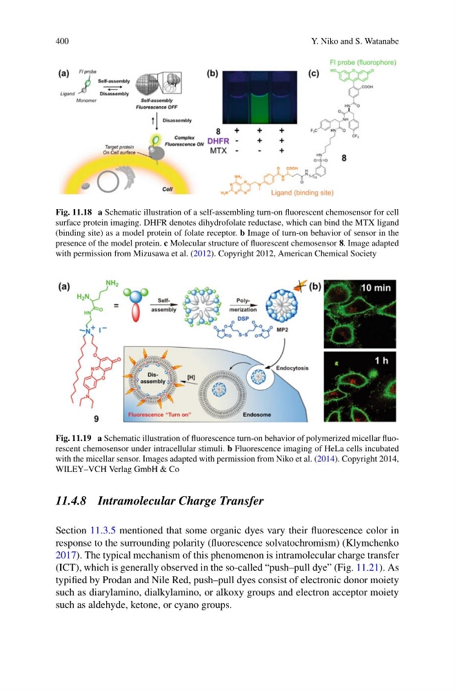
Fig. 11.18 a Schematic illustration of a self-assembling turn-on fluorescent chemosensor for cell
surface protein imaging. DHFR denotes dihydrofolate reductase, which can bind the MTX ligand
(binding site) as a model protein of folate receptor. b Image of turn-on behavior of sensor in the
presence of the model protein. c Molecular structure of fluorescent chemosensor 8. Image adapted
with permission from Mizusawa et al. (2012). Copyright 2012, American Chemical Society
Fig. 11.19 a Schematic illustration of fluorescence turn-on behavior of polymerized micellar fluorescent chemosensor under intracellular stimuli. b Fluorescence imaging of HeLa cells incubated
with the micellar sensor. Images adapted with permission from Niko et al. (2014). Copyright 2014,
WILEY–VCH Verlag GmbH & Co
11.4.8 Intramolecular Charge Transfer
Section 11.3.5 mentioned that some organic dyes vary their fluorescence color in
response to the surrounding polarity (fluorescence solvatochromism) (Klymchenko
2017). The typical mechanism of this phenomenon is intramolecular charge transfer
(ICT), which is generally observed in the so-called “push–pull dye” (Fig. 11.21). As
typified by Prodan and Nile Red, push–pull dyes consist of electronic donor moiety
such as diarylamino, dialkylamino, or alkoxy groups and electron acceptor moiety
such as aldehyde, ketone, or cyano groups.
Y. Niko and S. Watanabe
Fig. 11.18 a Schematic illustration of a self-assembling turn-on fluorescent chemosensor for cell
surface protein imaging. DHFR denotes dihydrofolate reductase, which can bind the MTX ligand
(binding site) as a model protein of folate receptor. b Image of turn-on behavior of sensor in the
presence of the model protein. c Molecular structure of fluorescent chemosensor 8. Image adapted
with permission from Mizusawa et al. (2012). Copyright 2012, American Chemical Society
Fig. 11.19 a Schematic illustration of fluorescence turn-on behavior of polymerized micellar fluorescent chemosensor under intracellular stimuli. b Fluorescence imaging of HeLa cells incubated
with the micellar sensor. Images adapted with permission from Niko et al. (2014). Copyright 2014,
WILEY–VCH Verlag GmbH & Co
11.4.8 Intramolecular Charge Transfer
Section 11.3.5 mentioned that some organic dyes vary their fluorescence color in
response to the surrounding polarity (fluorescence solvatochromism) (Klymchenko
2017). The typical mechanism of this phenomenon is intramolecular charge transfer
(ICT), which is generally observed in the so-called “push–pull dye” (Fig. 11.21). As
typified by Prodan and Nile Red, push–pull dyes consist of electronic donor moiety
such as diarylamino, dialkylamino, or alkoxy groups and electron acceptor moiety
such as aldehyde, ketone, or cyano groups.