

11 Fluorescent Chemosensors
399
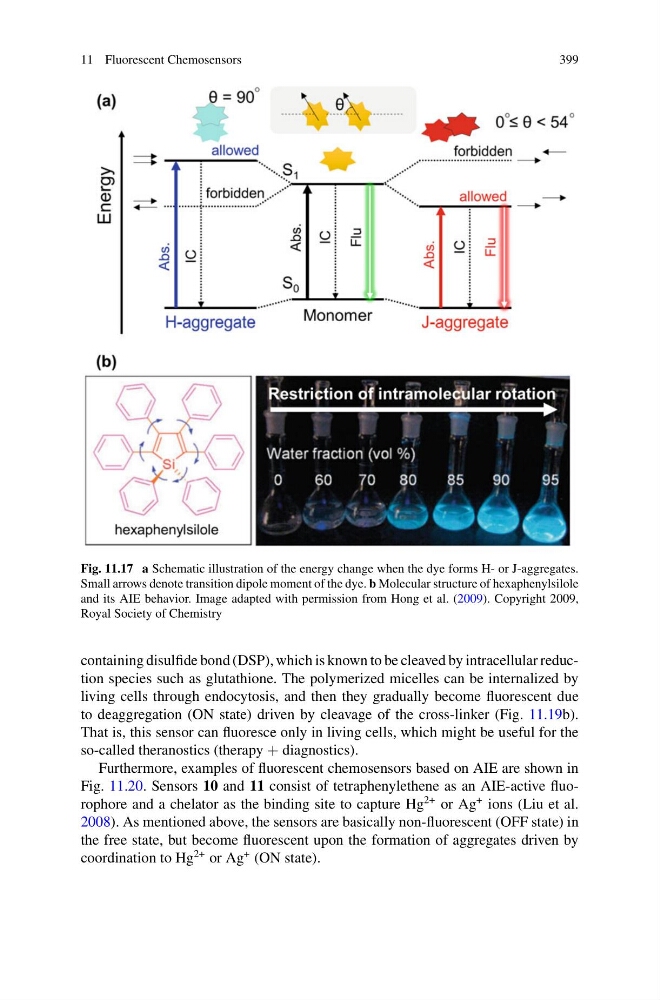
Fig. 11.17 a Schematic illustration of the energy change when the dye forms H- or J-aggregates.
Small arrows denote transition dipole moment of the dye. b Molecular structure of hexaphenylsilole
and its AIE behavior. Image adapted with permission from Hong et al. (2009). Copyright 2009,
Royal Society of Chemistry
containing disulfide bond (DSP), which is known to be cleaved by intracellular reduction species such as glutathione. The polymerized micelles can be internalized by
living cells through endocytosis, and then they gradually become fluorescent due
to deaggregation (ON state) driven by cleavage of the cross-linker (Fig. 11.19b).
That is, this sensor can fluoresce only in living cells, which might be useful for the
so-called theranostics (therapy + diagnostics).
Furthermore, examples of fluorescent chemosensors based on AIE are shown in
Fig. 11.20. Sensors 10 and 11 consist of tetraphenylethene as an AIE-active fluorophore and a chelator as the binding site to capture Hg
2+ or Ag
+ ions (Liu et al.
2008). As mentioned above, the sensors are basically non-fluorescent (OFF state) in
the free state, but become fluorescent upon the formation of aggregates driven by
coordination to Hg
2+ or Ag
+ (ON state).
399
Fig. 11.17 a Schematic illustration of the energy change when the dye forms H- or J-aggregates.
Small arrows denote transition dipole moment of the dye. b Molecular structure of hexaphenylsilole
and its AIE behavior. Image adapted with permission from Hong et al. (2009). Copyright 2009,
Royal Society of Chemistry
containing disulfide bond (DSP), which is known to be cleaved by intracellular reduction species such as glutathione. The polymerized micelles can be internalized by
living cells through endocytosis, and then they gradually become fluorescent due
to deaggregation (ON state) driven by cleavage of the cross-linker (Fig. 11.19b).
That is, this sensor can fluoresce only in living cells, which might be useful for the
so-called theranostics (therapy + diagnostics).
Furthermore, examples of fluorescent chemosensors based on AIE are shown in
Fig. 11.20. Sensors 10 and 11 consist of tetraphenylethene as an AIE-active fluorophore and a chelator as the binding site to capture Hg
2+ or Ag
+ ions (Liu et al.
2008). As mentioned above, the sensors are basically non-fluorescent (OFF state) in
the free state, but become fluorescent upon the formation of aggregates driven by
coordination to Hg
2+ or Ag
+ (ON state).