

11 Fluorescent Chemosensors
381
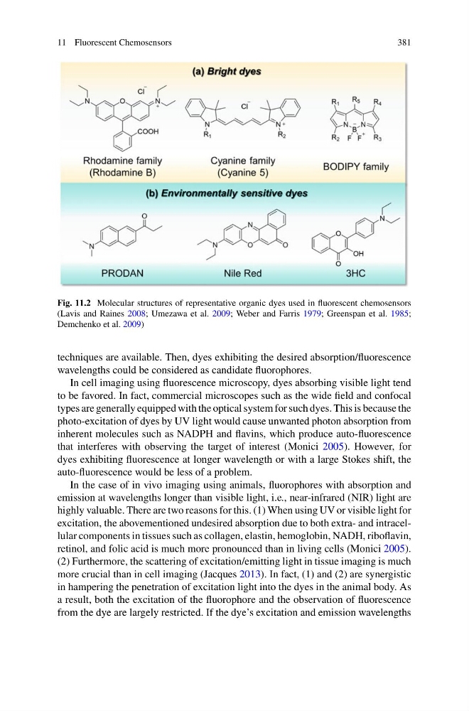
Fig. 11.2 Molecular structures of representative organic dyes used in fluorescent chemosensors
(Lavis and Raines 2008; Umezawa et al. 2009; Weber and Farris 1979; Greenspan et al. 1985;
Demchenko et al. 2009)
techniques are available. Then, dyes exhibiting the desired absorption/fluorescence
wavelengths could be considered as candidate fluorophores.
In cell imaging using fluorescence microscopy, dyes absorbing visible light tend
to be favored. In fact, commercial microscopes such as the wide field and confocal
types are generally equipped with the optical system for such dyes. This is because the
photo-excitation of dyes by UV light would cause unwanted photon absorption from
inherent molecules such as NADPH and flavins, which produce auto-fluorescence
that interferes with observing the target of interest (Monici 2005). However, for
dyes exhibiting fluorescence at longer wavelength or with a large Stokes shift, the
auto-fluorescence would be less of a problem.
In the case of in vivo imaging using animals, fluorophores with absorption and
emission at wavelengths longer than visible light, i.e., near-infrared (NIR) light are
highly valuable. There are two reasons for this. (1) When using UV or visible light for
excitation, the abovementioned undesired absorption due to both extra- and intracellular components in tissues such as collagen, elastin, hemoglobin, NADH, riboflavin,
retinol, and folic acid is much more pronounced than in living cells (Monici 2005).
(2) Furthermore, the scattering of excitation/emitting light in tissue imaging is much
more crucial than in cell imaging (Jacques 2013). In fact, (1) and (2) are synergistic
in hampering the penetration of excitation light into the dyes in the animal body. As
a result, both the excitation of the fluorophore and the observation of fluorescence
from the dye are largely restricted. If the dye’s excitation and emission wavelengths
381
Fig. 11.2 Molecular structures of representative organic dyes used in fluorescent chemosensors
(Lavis and Raines 2008; Umezawa et al. 2009; Weber and Farris 1979; Greenspan et al. 1985;
Demchenko et al. 2009)
techniques are available. Then, dyes exhibiting the desired absorption/fluorescence
wavelengths could be considered as candidate fluorophores.
In cell imaging using fluorescence microscopy, dyes absorbing visible light tend
to be favored. In fact, commercial microscopes such as the wide field and confocal
types are generally equipped with the optical system for such dyes. This is because the
photo-excitation of dyes by UV light would cause unwanted photon absorption from
inherent molecules such as NADPH and flavins, which produce auto-fluorescence
that interferes with observing the target of interest (Monici 2005). However, for
dyes exhibiting fluorescence at longer wavelength or with a large Stokes shift, the
auto-fluorescence would be less of a problem.
In the case of in vivo imaging using animals, fluorophores with absorption and
emission at wavelengths longer than visible light, i.e., near-infrared (NIR) light are
highly valuable. There are two reasons for this. (1) When using UV or visible light for
excitation, the abovementioned undesired absorption due to both extra- and intracellular components in tissues such as collagen, elastin, hemoglobin, NADH, riboflavin,
retinol, and folic acid is much more pronounced than in living cells (Monici 2005).
(2) Furthermore, the scattering of excitation/emitting light in tissue imaging is much
more crucial than in cell imaging (Jacques 2013). In fact, (1) and (2) are synergistic
in hampering the penetration of excitation light into the dyes in the animal body. As
a result, both the excitation of the fluorophore and the observation of fluorescence
from the dye are largely restricted. If the dye’s excitation and emission wavelengths