

300
T. Ishi-i
dye
DSPE-PEG 2000
CH3(CH2)16
O
O
O
O
P
O
O
CH3(CH2)16
O
N
(OCH2CH2)45OCH3
O
H
O -
NH4 +
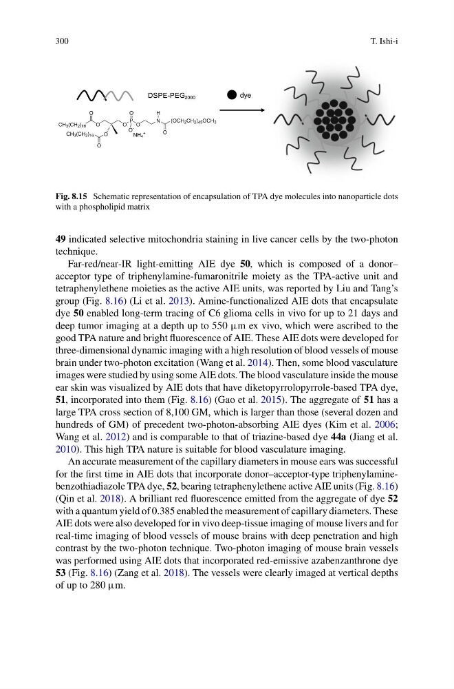
Fig. 8.15 Schematic representation of encapsulation of TPA dye molecules into nanoparticle dots
with a phospholipid matrix
49 indicated selective mitochondria staining in live cancer cells by the two-photon
technique.
Far-red/near-IR light-emitting AIE dye 50, which is composed of a donor–
acceptor type of triphenylamine-fumaronitrile moiety as the TPA-active unit and
tetraphenylethene moieties as the active AIE units, was reported by Liu and Tang’s
group (Fig. 8.16) (Li et al. 2013). Amine-functionalized AIE dots that encapsulate
dye 50 enabled long-term tracing of C6 glioma cells in vivo for up to 21 days and
deep tumor imaging at a depth up to 550 μm ex vivo, which were ascribed to the
good TPA nature and bright fluorescence of AIE. These AIE dots were developed for
three-dimensional dynamic imaging with a high resolution of blood vessels of mouse
brain under two-photon excitation (Wang et al. 2014). Then, some blood vasculature
images were studied by using some AIE dots. The blood vasculature inside the mouse
ear skin was visualized by AIE dots that have diketopyrrolopyrrole-based TPA dye,
51, incorporated into them (Fig. 8.16) (Gao et al. 2015). The aggregate of 51 has a
large TPA cross section of 8,100 GM, which is larger than those (several dozen and
hundreds of GM) of precedent two-photon-absorbing AIE dyes (Kim et al. 2006;
Wang et al. 2012) and is comparable to that of triazine-based dye 44a (Jiang et al.
2010). This high TPA nature is suitable for blood vasculature imaging.
An accurate measurement of the capillary diameters in mouse ears was successful
for the first time in AIE dots that incorporate donor–acceptor-type triphenylaminebenzothiadiazole TPA dye, 52, bearing tetraphenylethene active AIE units (Fig. 8.16)
(Qin et al. 2018). A brilliant red fluorescence emitted from the aggregate of dye 52
with a quantum yield of 0.385 enabled the measurement of capillary diameters. These
AIE dots were also developed for in vivo deep-tissue imaging of mouse livers and for
real-time imaging of blood vessels of mouse brains with deep penetration and high
contrast by the two-photon technique. Two-photon imaging of mouse brain vessels
was performed using AIE dots that incorporated red-emissive azabenzanthrone dye
53 (Fig. 8.16) (Zang et al. 2018). The vessels were clearly imaged at vertical depths
of up to 280 μm.
T. Ishi-i
dye
DSPE-PEG 2000
CH3(CH2)16
O
O
O
O
P
O
O
CH3(CH2)16
O
N
(OCH2CH2)45OCH3
O
H
O -
NH4 +
Fig. 8.15 Schematic representation of encapsulation of TPA dye molecules into nanoparticle dots
with a phospholipid matrix
49 indicated selective mitochondria staining in live cancer cells by the two-photon
technique.
Far-red/near-IR light-emitting AIE dye 50, which is composed of a donor–
acceptor type of triphenylamine-fumaronitrile moiety as the TPA-active unit and
tetraphenylethene moieties as the active AIE units, was reported by Liu and Tang’s
group (Fig. 8.16) (Li et al. 2013). Amine-functionalized AIE dots that encapsulate
dye 50 enabled long-term tracing of C6 glioma cells in vivo for up to 21 days and
deep tumor imaging at a depth up to 550 μm ex vivo, which were ascribed to the
good TPA nature and bright fluorescence of AIE. These AIE dots were developed for
three-dimensional dynamic imaging with a high resolution of blood vessels of mouse
brain under two-photon excitation (Wang et al. 2014). Then, some blood vasculature
images were studied by using some AIE dots. The blood vasculature inside the mouse
ear skin was visualized by AIE dots that have diketopyrrolopyrrole-based TPA dye,
51, incorporated into them (Fig. 8.16) (Gao et al. 2015). The aggregate of 51 has a
large TPA cross section of 8,100 GM, which is larger than those (several dozen and
hundreds of GM) of precedent two-photon-absorbing AIE dyes (Kim et al. 2006;
Wang et al. 2012) and is comparable to that of triazine-based dye 44a (Jiang et al.
2010). This high TPA nature is suitable for blood vasculature imaging.
An accurate measurement of the capillary diameters in mouse ears was successful
for the first time in AIE dots that incorporate donor–acceptor-type triphenylaminebenzothiadiazole TPA dye, 52, bearing tetraphenylethene active AIE units (Fig. 8.16)
(Qin et al. 2018). A brilliant red fluorescence emitted from the aggregate of dye 52
with a quantum yield of 0.385 enabled the measurement of capillary diameters. These
AIE dots were also developed for in vivo deep-tissue imaging of mouse livers and for
real-time imaging of blood vessels of mouse brains with deep penetration and high
contrast by the two-photon technique. Two-photon imaging of mouse brain vessels
was performed using AIE dots that incorporated red-emissive azabenzanthrone dye
53 (Fig. 8.16) (Zang et al. 2018). The vessels were clearly imaged at vertical depths
of up to 280 μm.