

10.7.5 Arrays of supported bilayers and microfluidic
platforms
Spatially addressed microarrays have found use as extremely rapid and
powerful means of data collection. Unlike DNA-, protein-, or peptidebased arrays, phospholipid bilayer systems must remain hydrated at all
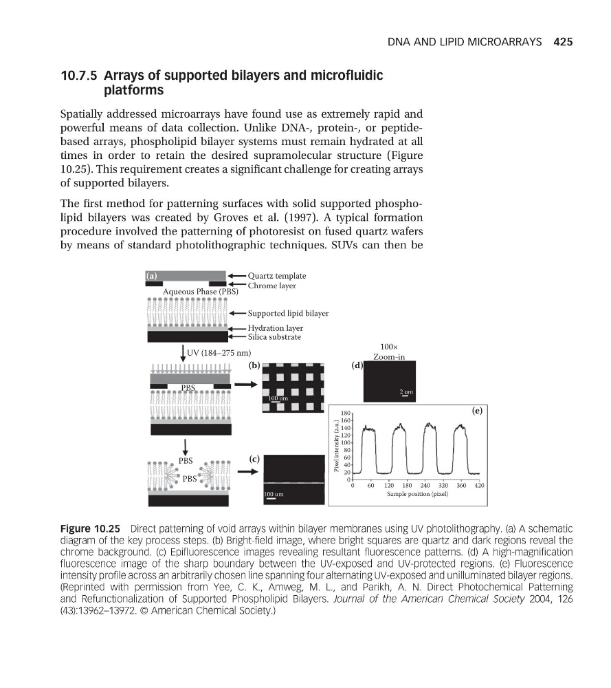
times in order to retain the desired supramolecular structure (Figure
10.25). This requirement creates a significant challenge for creating arrays
of supported bilayers.
The first method for patterning surfaces with solid supported phospholipid bilayers was created by Groves et al. (1997). A typical formation
procedure involved the patterning of photoresist on fused quartz wafers
by means of standard photolithographic techniques. SUVs can then be
(a)
(b)
100×
Zoom-in
(c)
180
160
140
120
100
80
60
40
20
Pixel intensity (a.u.)
0
0
60
120 180
Sample position (pixel)
240 320 360 420
(d)
(e)
100 um
100 um
2 um
Quartz template
Aqueous Phase (PBS)
Chrome layer
Supported lipid bilayer
Silica substrate
UV (184–275 nm)
PBS
PBS
PBS
Hydration layer
Figure 10.25 Direct patterning of void arrays within bilayer membranes using UV photolithography. (a) A schematic
diagram of the key process steps. (b) Bright-field image, where bright squares are quartz and dark regions reveal the
chrome background. (c) Epifluorescence images revealing resultant fluorescence patterns. (d) A high-magnification
fluorescence image of the sharp boundary between the UV-exposed and UV-protected regions. (e) Fluorescence
intensity profile across an arbitrarily chosen line spanning four alternating UV-exposed and unilluminated bilayer regions.
(Reprinted with permission from Yee, C. K., Amweg, M. L., and Parikh, A. N. Direct Photochemical Patterning
and Refunctionalization of Supported Phospholipid Bilayers. Journal of the American Chemical Society 2004, 126
(43):13962–13972. © American Chemical Society.)
DNA AND LIPID MICROARRAYS 425
platforms
Spatially addressed microarrays have found use as extremely rapid and
powerful means of data collection. Unlike DNA-, protein-, or peptidebased arrays, phospholipid bilayer systems must remain hydrated at all
times in order to retain the desired supramolecular structure (Figure
10.25). This requirement creates a significant challenge for creating arrays
of supported bilayers.
The first method for patterning surfaces with solid supported phospholipid bilayers was created by Groves et al. (1997). A typical formation
procedure involved the patterning of photoresist on fused quartz wafers
by means of standard photolithographic techniques. SUVs can then be
(a)
(b)
100×
Zoom-in
(c)
180
160
140
120
100
80
60
40
20
Pixel intensity (a.u.)
0
0
60
120 180
Sample position (pixel)
240 320 360 420
(d)
(e)
100 um
100 um
2 um
Quartz template
Aqueous Phase (PBS)
Chrome layer
Supported lipid bilayer
Silica substrate
UV (184–275 nm)
PBS
PBS
PBS
Hydration layer
Figure 10.25 Direct patterning of void arrays within bilayer membranes using UV photolithography. (a) A schematic
diagram of the key process steps. (b) Bright-field image, where bright squares are quartz and dark regions reveal the
chrome background. (c) Epifluorescence images revealing resultant fluorescence patterns. (d) A high-magnification
fluorescence image of the sharp boundary between the UV-exposed and UV-protected regions. (e) Fluorescence
intensity profile across an arbitrarily chosen line spanning four alternating UV-exposed and unilluminated bilayer regions.
(Reprinted with permission from Yee, C. K., Amweg, M. L., and Parikh, A. N. Direct Photochemical Patterning
and Refunctionalization of Supported Phospholipid Bilayers. Journal of the American Chemical Society 2004, 126
(43):13962–13972. © American Chemical Society.)
DNA AND LIPID MICROARRAYS 425