

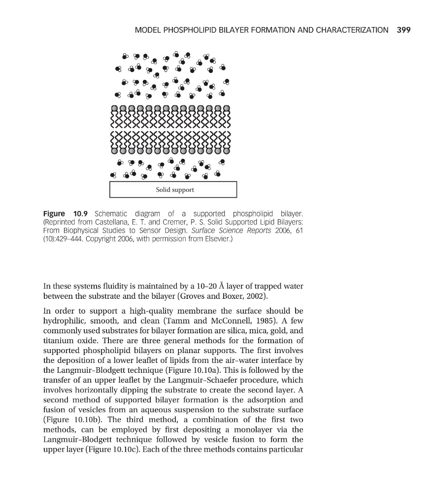
In these systems fluidity is maintained by a 10–20 Å layer of trapped water
between the substrate and the bilayer (Groves and Boxer, 2002).
In order to support a high-quality membrane the surface should be
hydrophilic, smooth, and clean (Tamm and McConnell, 1985). A few
commonly used substrates for bilayer formation are silica, mica, gold, and
titanium oxide. There are three general methods for the formation of
supported phospholipid bilayers on planar supports. The first involves
the deposition of a lower leaflet of lipids from the air–water interface by
the Langmuir–Blodgett technique (Figure 10.10a). This is followed by the
transfer of an upper leaflet by the Langmuir–Schaefer procedure, which
involves horizontally dipping the substrate to create the second layer. A
second method of supported bilayer formation is the adsorption and
fusion of vesicles from an aqueous suspension to the substrate surface
(Figure 10.10b). The third method, a combination of the first two
methods, can be employed by first depositing a monolayer via the
Langmuir–Blodgett technique followed by vesicle fusion to form the
upper layer (Figure 10.10c). Each of the three methods contains particular
Solid support
Figure 10.9 Schematic diagram of a supported phospholipid bilayer.
(Reprinted from Castellana, E. T. and Cremer, P. S. Solid Supported Lipid Bilayers:
From Biophysical Studies to Sensor Design. Surface Science Reports 2006, 61
(10):429–444. Copyright 2006, with permission from Elsevier.)
MODEL PHOSPHOLIPID BILAYER FORMATION AND CHARACTERIZATION 399
between the substrate and the bilayer (Groves and Boxer, 2002).
In order to support a high-quality membrane the surface should be
hydrophilic, smooth, and clean (Tamm and McConnell, 1985). A few
commonly used substrates for bilayer formation are silica, mica, gold, and
titanium oxide. There are three general methods for the formation of
supported phospholipid bilayers on planar supports. The first involves
the deposition of a lower leaflet of lipids from the air–water interface by
the Langmuir–Blodgett technique (Figure 10.10a). This is followed by the
transfer of an upper leaflet by the Langmuir–Schaefer procedure, which
involves horizontally dipping the substrate to create the second layer. A
second method of supported bilayer formation is the adsorption and
fusion of vesicles from an aqueous suspension to the substrate surface
(Figure 10.10b). The third method, a combination of the first two
methods, can be employed by first depositing a monolayer via the
Langmuir–Blodgett technique followed by vesicle fusion to form the
upper layer (Figure 10.10c). Each of the three methods contains particular
Solid support
Figure 10.9 Schematic diagram of a supported phospholipid bilayer.
(Reprinted from Castellana, E. T. and Cremer, P. S. Solid Supported Lipid Bilayers:
From Biophysical Studies to Sensor Design. Surface Science Reports 2006, 61
(10):429–444. Copyright 2006, with permission from Elsevier.)
MODEL PHOSPHOLIPID BILAYER FORMATION AND CHARACTERIZATION 399