

silicon or another metal and then shining the laser through this nanometric aperture. Both of these probes can be manufactured with aperture
diameters of ~50–100 nm. Recent developments have also been made in
so-called apertureless NSOM probes. These probes exhibit great promise
in achieving even better subdiffraction limit resolutions.
An NSOM setup can be operated in at least five different modes, as listed
below (see Figure 8.37).
1. Transmission mode. Light is shone through the NSOM probe and then
detected on the other side of the sample.
2. Reflection mode. Light is passed through the NSOM probe and
reflected from the sample. This reflected light is detected and compiled to produce an image.
3. Transmission-collection mode. Light is shone on the sample from
underneath and the transmitted light is collected in the NSOM probe
and passed to a detector.
4. Reflection-collection mode. Light is shone on the sample from above
and reflected light is collected by the NSOM probe and sent to a
detector.
5. Illumination-collection mode. The NSOM probe is used as both the
light source and the light collector.
200 nm
200 nm
(a)
(b)
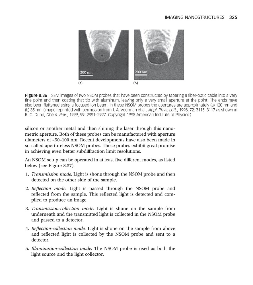
Figure 8.36 SEM images of two NSOM probes that have been constructed by tapering a fiber-optic cable into a very
fine point and then coating that tip with aluminum, leaving only a very small aperture at the point. The ends have
also been flattened using a focused ion beam. In these NSOM probes the apertures are approximately (a) 120 nm and
(b) 35 nm. (Image reprinted with permission from J. A. Veerman et al., Appl. Phys. Lett., 1998, 72: 3115–3117 as shown in
R. C. Dunn, Chem. Rev., 1999, 99: 2891–2927. Copyright 1998 American Institute of Physics.)
IMAGING NANOSTRUCTURES 325
diameters of ~50–100 nm. Recent developments have also been made in
so-called apertureless NSOM probes. These probes exhibit great promise
in achieving even better subdiffraction limit resolutions.
An NSOM setup can be operated in at least five different modes, as listed
below (see Figure 8.37).
1. Transmission mode. Light is shone through the NSOM probe and then
detected on the other side of the sample.
2. Reflection mode. Light is passed through the NSOM probe and
reflected from the sample. This reflected light is detected and compiled to produce an image.
3. Transmission-collection mode. Light is shone on the sample from
underneath and the transmitted light is collected in the NSOM probe
and passed to a detector.
4. Reflection-collection mode. Light is shone on the sample from above
and reflected light is collected by the NSOM probe and sent to a
detector.
5. Illumination-collection mode. The NSOM probe is used as both the
light source and the light collector.
200 nm
200 nm
(a)
(b)
Figure 8.36 SEM images of two NSOM probes that have been constructed by tapering a fiber-optic cable into a very
fine point and then coating that tip with aluminum, leaving only a very small aperture at the point. The ends have
also been flattened using a focused ion beam. In these NSOM probes the apertures are approximately (a) 120 nm and
(b) 35 nm. (Image reprinted with permission from J. A. Veerman et al., Appl. Phys. Lett., 1998, 72: 3115–3117 as shown in
R. C. Dunn, Chem. Rev., 1999, 99: 2891–2927. Copyright 1998 American Institute of Physics.)
IMAGING NANOSTRUCTURES 325