

Scanning electron microscopy (SEM) is a commonly used electron
microscopy method that functions by monitoring the electrons that are
backscattered after interacting with a sample and uses those backscattered electrons to reconstruct an image of the sample. On the other hand,
transmission electron microscopy (TEM) makes use of electrons that
20 nm
20 nm
(a)
(b)
(c)
1 µm
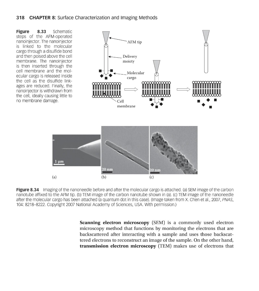
Figure 8.34 Imaging of the nanoneedle before and after the molecular cargo is attached. (a) SEM image of the carbon
nanotube affixed to the AFM tip. (b) TEM image of the carbon nanotube shown in (a). (c) TEM image of the nanoneedle
after the molecular cargo has been attached (a quantum dot in this case). (Image taken from X. Chen et al., 2007, PNAS,
104: 8218–8222. Copyright 2007 National Academy of Sciences, USA. With permission.)
AFM tip
Delivery
moiety
Molecular
cargo
Cell
membrane
Figure
8.33
Schematic
steps of the AFM-operated
nanoinjector. The nanoinjector
is linked to the molecular
cargo through a disulfide bond
and then poised above the cell
membrane. The nanoinjector
is then inserted through the
cell membrane and the molecular cargo is released inside
the cell as the disulfide linkages are reduced. Finally, the
nanoinjector is withdrawn from
the cell, ideally causing little to
no membrane damage.
CHAPTER 8: Surface Characterization and Imaging Methods
318
microscopy method that functions by monitoring the electrons that are
backscattered after interacting with a sample and uses those backscattered electrons to reconstruct an image of the sample. On the other hand,
transmission electron microscopy (TEM) makes use of electrons that
20 nm
20 nm
(a)
(b)
(c)
1 µm
Figure 8.34 Imaging of the nanoneedle before and after the molecular cargo is attached. (a) SEM image of the carbon
nanotube affixed to the AFM tip. (b) TEM image of the carbon nanotube shown in (a). (c) TEM image of the nanoneedle
after the molecular cargo has been attached (a quantum dot in this case). (Image taken from X. Chen et al., 2007, PNAS,
104: 8218–8222. Copyright 2007 National Academy of Sciences, USA. With permission.)
AFM tip
Delivery
moiety
Molecular
cargo
Cell
membrane
Figure
8.33
Schematic
steps of the AFM-operated
nanoinjector. The nanoinjector
is linked to the molecular
cargo through a disulfide bond
and then poised above the cell
membrane. The nanoinjector
is then inserted through the
cell membrane and the molecular cargo is released inside
the cell as the disulfide linkages are reduced. Finally, the
nanoinjector is withdrawn from
the cell, ideally causing little to
no membrane damage.
CHAPTER 8: Surface Characterization and Imaging Methods
318