

such as fluorescence recovery after photobleaching (FRAP), fluorescence
resonant energy transfer (FRET), and fluorescence interference contrast
microscopy (FLIC), which are discussed in Chapters 9 and 10. X-ray
fluorescence is also commonly used in dyes. These fluorescing dyes can
be attached to target molecules of interest that can be excited and
observed in conditions where dyes in the visual wavelengths of light are
not usable. This is often the case for in vivo studies when there is a layer
between the observer and the dyed molecule that allows for x-rays to pass
through but does not allow visible light to pass through.
6.3.3 Diffraction
When x-rays pass through matter, the radiation interacts with electrons in
the matter in such a way that the path of the x-rays can be altered. This
scattering effect of matter on x-rays is known as diffraction. In a crystal or
any ordered sample, the x-rays scatter in ways that produce higherintensity areas and lower-intensity areas, also termed constructive and
destructive interference, respectively. By comparing these high- and
low-intensity areas, it is possible to determine the architecture and the
ordering in matter at the nanoscale. It is, of course, slightly more complicated in practice, because when an x-ray beam strikes an ordered
crystal, which can be thought of as multiple layers of atoms, each subsequent layer of atoms scatters some of the beam and lets the remainder
of the beam through. So that this diffraction can take place in a way that
allows us to relate it to the materials structure, the space between layers of
atoms in the material must be about the same distance as the wavelength
of the radiation used to probe it and the atoms comprising the system
must be highly ordered with few defects.
In order to use the high- and low-intensity areas that result from shining x-rays through an ordered crystal, a way to relate them to the structure must be known. Fortunately, in 1912, W. L. Bragg determined this
relationship. The Bragg equation for constructive interference is as
follows:
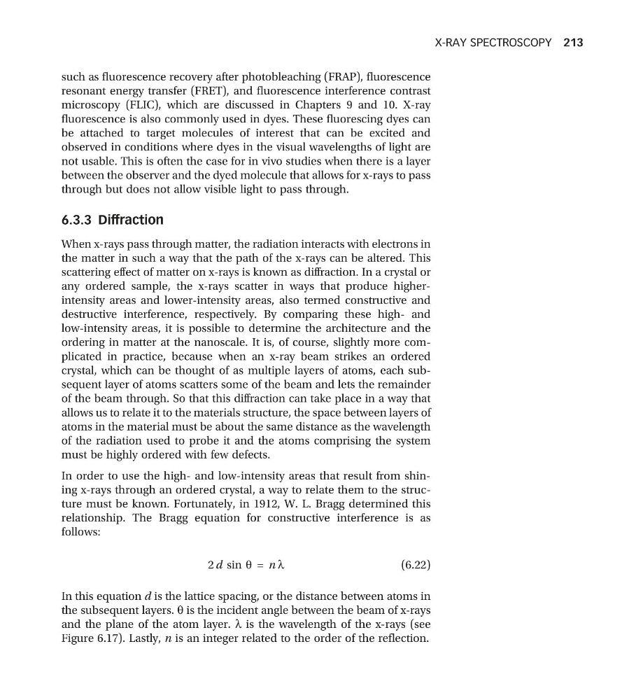
2 d sin q = n l
(6.22)
In this equation d is the lattice spacing, or the distance between atoms in
the subsequent layers. q is the incident angle between the beam of x-rays
and the plane of the atom layer. l is the wavelength of the x-rays (see
Figure 6.17). Lastly, n is an integer related to the order of the reflection.
X-RAY SPECTROSCOPY 213
resonant energy transfer (FRET), and fluorescence interference contrast
microscopy (FLIC), which are discussed in Chapters 9 and 10. X-ray
fluorescence is also commonly used in dyes. These fluorescing dyes can
be attached to target molecules of interest that can be excited and
observed in conditions where dyes in the visual wavelengths of light are
not usable. This is often the case for in vivo studies when there is a layer
between the observer and the dyed molecule that allows for x-rays to pass
through but does not allow visible light to pass through.
6.3.3 Diffraction
When x-rays pass through matter, the radiation interacts with electrons in
the matter in such a way that the path of the x-rays can be altered. This
scattering effect of matter on x-rays is known as diffraction. In a crystal or
any ordered sample, the x-rays scatter in ways that produce higherintensity areas and lower-intensity areas, also termed constructive and
destructive interference, respectively. By comparing these high- and
low-intensity areas, it is possible to determine the architecture and the
ordering in matter at the nanoscale. It is, of course, slightly more complicated in practice, because when an x-ray beam strikes an ordered
crystal, which can be thought of as multiple layers of atoms, each subsequent layer of atoms scatters some of the beam and lets the remainder
of the beam through. So that this diffraction can take place in a way that
allows us to relate it to the materials structure, the space between layers of
atoms in the material must be about the same distance as the wavelength
of the radiation used to probe it and the atoms comprising the system
must be highly ordered with few defects.
In order to use the high- and low-intensity areas that result from shining x-rays through an ordered crystal, a way to relate them to the structure must be known. Fortunately, in 1912, W. L. Bragg determined this
relationship. The Bragg equation for constructive interference is as
follows:
2 d sin q = n l
(6.22)
In this equation d is the lattice spacing, or the distance between atoms in
the subsequent layers. q is the incident angle between the beam of x-rays
and the plane of the atom layer. l is the wavelength of the x-rays (see
Figure 6.17). Lastly, n is an integer related to the order of the reflection.
X-RAY SPECTROSCOPY 213