

Conventional, Molecular, and Biosensor-Based Detection Methods ◾ 559
mammalian cells as a result of exposure to an “external” or environmental challenge, such as the
presence of pathogens or toxins in clinical, environmental, or food samples, including Lis. monocytogenes and Esc. coli and toxins. Some of the cell-based assays also utilize the metabolic responses
of cells (e.g., cyanobacteria) to detect biological products, like oxygen and herbicides in water.
A novel B cell-based sensor was genetically engineered to express cytosolic aequorin, a calcium-sensitive bioluminescent protein, and pathogen-specific surface antibodies. When a pathogen binds to the surface antibodies, it generates a specific bioluminescent signal, and it has been
shown to be able to detect very low numbers of pathogenic bacteria, like Esc. coli O157:H7, Bacillus
anthracis spores, and Yersinia pestis in minutes. Artificial cells, also known as “liposomes,” made
up of a lipid bilayer with specific receptors carrying a fluorescent-tagged reporter molecule inside,
can serve as a sensitive sensor for detection of spore-forming toxins. Toxins first bind to a receptor and then disrupt the membrane, allowing the release of the fluorescent reporter, which could
be detected by a spectrofluorometer. The liposome can be stored in ambient conditions for up to
five months without the requirement for a cell culture facility, and it is able to detect nanogram
quantities of toxin in 30 minutes, and moreover this system can eliminate the requirement for a
cell culture facility. Mammalian cell-based biochips have also been used for detection of allergens
and viruses. For example, the cultured basophilic mast cell line (RBL-2H3), on interaction with
an allergen, releases a fluorescent-bound histamine, which is measured by a photomultiplier tube
attached to a microscope.
Conclusion
Microbial culturing methods are irreplaceable for microbiological analysis of food to monitor
product quality and safety. Advances in molecular methods have benefited pathogen detection
and identification in foods; however, these methods still lag behind for analysis of spoilagecausing microorganisms to assess shelf life or fermentative microorganisms in finished products.
Biosensor-based methods are in the early stage of their development; some of them are proven to
be fast and sensitive and have the potential to be built as an automated hand-held device for onsite
Pathogens or toxins
Enzyme
Receptor
Color,
impedance
Substrate
Interaction or
pathogens with
mammalian cells
on sensor
Cell lysis or release
of signaling
molecules
Detection
using a
sensor
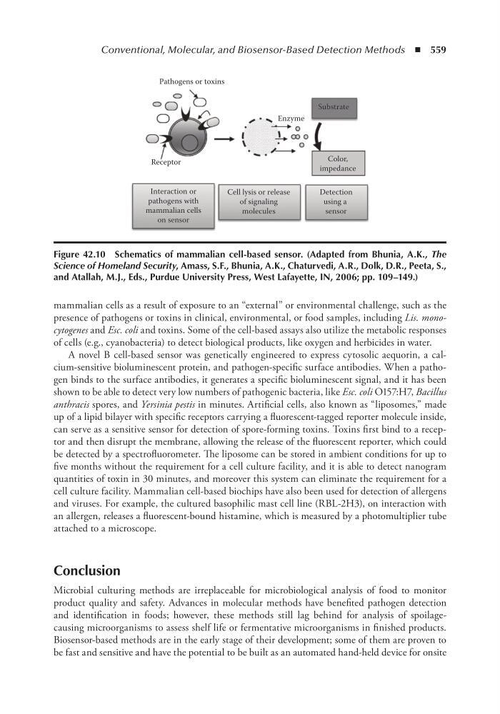
Figure 42.10 Schematics of mammalian cell-based sensor. (Adapted from Bhunia, A.K., The
Science of Homeland Security, Amass, S.F., Bhunia, A.K., Chaturvedi, A.R., Dolk, D.R., Peeta, S.,
and Atallah, M.J., eds., Purdue University Press, West Lafayette, in, 2006; pp. 109–149.)
mammalian cells as a result of exposure to an “external” or environmental challenge, such as the
presence of pathogens or toxins in clinical, environmental, or food samples, including Lis. monocytogenes and Esc. coli and toxins. Some of the cell-based assays also utilize the metabolic responses
of cells (e.g., cyanobacteria) to detect biological products, like oxygen and herbicides in water.
A novel B cell-based sensor was genetically engineered to express cytosolic aequorin, a calcium-sensitive bioluminescent protein, and pathogen-specific surface antibodies. When a pathogen binds to the surface antibodies, it generates a specific bioluminescent signal, and it has been
shown to be able to detect very low numbers of pathogenic bacteria, like Esc. coli O157:H7, Bacillus
anthracis spores, and Yersinia pestis in minutes. Artificial cells, also known as “liposomes,” made
up of a lipid bilayer with specific receptors carrying a fluorescent-tagged reporter molecule inside,
can serve as a sensitive sensor for detection of spore-forming toxins. Toxins first bind to a receptor and then disrupt the membrane, allowing the release of the fluorescent reporter, which could
be detected by a spectrofluorometer. The liposome can be stored in ambient conditions for up to
five months without the requirement for a cell culture facility, and it is able to detect nanogram
quantities of toxin in 30 minutes, and moreover this system can eliminate the requirement for a
cell culture facility. Mammalian cell-based biochips have also been used for detection of allergens
and viruses. For example, the cultured basophilic mast cell line (RBL-2H3), on interaction with
an allergen, releases a fluorescent-bound histamine, which is measured by a photomultiplier tube
attached to a microscope.
Conclusion
Microbial culturing methods are irreplaceable for microbiological analysis of food to monitor
product quality and safety. Advances in molecular methods have benefited pathogen detection
and identification in foods; however, these methods still lag behind for analysis of spoilagecausing microorganisms to assess shelf life or fermentative microorganisms in finished products.
Biosensor-based methods are in the early stage of their development; some of them are proven to
be fast and sensitive and have the potential to be built as an automated hand-held device for onsite
Pathogens or toxins
Enzyme
Receptor
Color,
impedance
Substrate
Interaction or
pathogens with
mammalian cells
on sensor
Cell lysis or release
of signaling
molecules
Detection
using a
sensor
Figure 42.10 Schematics of mammalian cell-based sensor. (Adapted from Bhunia, A.K., The
Science of Homeland Security, Amass, S.F., Bhunia, A.K., Chaturvedi, A.R., Dolk, D.R., Peeta, S.,
and Atallah, M.J., eds., Purdue University Press, West Lafayette, in, 2006; pp. 109–149.)