

Conventional, Molecular, and Biosensor-Based Detection Methods ◾ 547
containing the immobilized antigen. In this method, only the free-unbound antibody will bind
the immobilized antigen. After washing, a secondary antibody-enzyme conjugate and substrate
system are added for color development. The highest dilution of cells showing the minimum reaction, or equivalent to a background control, is considered positive. Generally, the detection limit
for all ELISA assays ranges from 10 6 to 10 7 bacterial cells/mL. The presence of that high level of
contaminants in a ready-to-eat product is not expected; therefore, an intermediate enrichment
step is necessary to increase cell population. Several commercial ELISA assay kits are currently
available for detection of foodborne pathogens from enriched food samples. 3,12
Immunofluorescence Assay
In principle, an immunofluorescence (IF) assay is similar to an ELISA assay. In IF, a fluorescently
labeled detection antibody (against somatic or flagellar antigens of a pathogen) that emits fluorescence after forming a complex with an antigen on a glass slide or in a 96-well microtiter plate is
used, which is detected by a fluorescence microscope, a digital camera, or a spectrofluorometer.
The fluorescent markers used are rhodamine B, fluorescein isocyanate, and fluorescein isothiocyanate (FITC). The fluorescent antibody technique can be carried out by using two basic methods:
The direct method employs binding of antigens with a fluorescent-labeled specific antibody. In
the indirect method, the primary antibody is not coupled with the fluorescence, but instead, a
species-specific secondary antibody is labeled with fluorescence. In the indirect method, the labeled
antibody detects the presence of the primary antibody and antigen complex. The use of the indirect
method eliminates the need to prepare a fluorescent-labeled antibody for each organism of interest.
Immunochromatographic Lateral Flow Assay
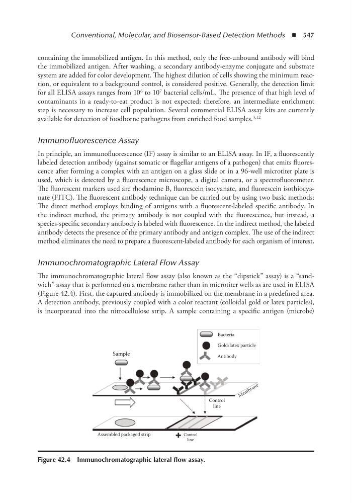
The immunochromatographic lateral flow assay (also known as the “dipstick” assay) is a “sandwich” assay that is performed on a membrane rather than in microtiter wells as are used in ELISA
(Figure 42.4). First, the captured antibody is immobilized on the membrane in a predefined area.
A detection antibody, previously coupled with a color reactant (colloidal gold or latex particles),
is incorporated into the nitrocellulose strip. A sample containing a specific antigen (microbe)
Bacteria
Gold/latex particle
Antibody
Sample
Control
line
M e m b r a n e
Assembled packaged strip
Control
line
Figure 42.4 immunochromatographic lateral flow assay.
containing the immobilized antigen. In this method, only the free-unbound antibody will bind
the immobilized antigen. After washing, a secondary antibody-enzyme conjugate and substrate
system are added for color development. The highest dilution of cells showing the minimum reaction, or equivalent to a background control, is considered positive. Generally, the detection limit
for all ELISA assays ranges from 10 6 to 10 7 bacterial cells/mL. The presence of that high level of
contaminants in a ready-to-eat product is not expected; therefore, an intermediate enrichment
step is necessary to increase cell population. Several commercial ELISA assay kits are currently
available for detection of foodborne pathogens from enriched food samples. 3,12
Immunofluorescence Assay
In principle, an immunofluorescence (IF) assay is similar to an ELISA assay. In IF, a fluorescently
labeled detection antibody (against somatic or flagellar antigens of a pathogen) that emits fluorescence after forming a complex with an antigen on a glass slide or in a 96-well microtiter plate is
used, which is detected by a fluorescence microscope, a digital camera, or a spectrofluorometer.
The fluorescent markers used are rhodamine B, fluorescein isocyanate, and fluorescein isothiocyanate (FITC). The fluorescent antibody technique can be carried out by using two basic methods:
The direct method employs binding of antigens with a fluorescent-labeled specific antibody. In
the indirect method, the primary antibody is not coupled with the fluorescence, but instead, a
species-specific secondary antibody is labeled with fluorescence. In the indirect method, the labeled
antibody detects the presence of the primary antibody and antigen complex. The use of the indirect
method eliminates the need to prepare a fluorescent-labeled antibody for each organism of interest.
Immunochromatographic Lateral Flow Assay
The immunochromatographic lateral flow assay (also known as the “dipstick” assay) is a “sandwich” assay that is performed on a membrane rather than in microtiter wells as are used in ELISA
(Figure 42.4). First, the captured antibody is immobilized on the membrane in a predefined area.
A detection antibody, previously coupled with a color reactant (colloidal gold or latex particles),
is incorporated into the nitrocellulose strip. A sample containing a specific antigen (microbe)
Bacteria
Gold/latex particle
Antibody
Sample
Control
line
M e m b r a n e
Assembled packaged strip
Control
line
Figure 42.4 immunochromatographic lateral flow assay.