

Conventional, Molecular, and Biosensor-Based Detection Methods ◾ 545
they only exhibit magnetic properties in the presence of an external magnetic field. The most common magnetic carriers are the Dynabeads ® with diameter ranging from 1 to 2.8 μm. These are
polystyrene-coated iron oxide. The streptavidin-coated beads are available for immobilization of a
biotinylated antibody. They can be easily removed from a suspension by using a magnetic separator. Because there is no magnetic remnant, the particles are not attracted to each other and therefore can be easily suspended in a homogeneous mixture in the absence of any external magnetic
field. IMS has two steps: First, the suspension containing the microbes of interest is mixed with
magnetic particles for about 30–60 minutes, and then the tube containing the magnetic complex
is placed in a magnetic separator, and the liquid is poured off. Second, the magnetic complex is
washed several times with a buffer to remove unwanted contaminants, and then the cells are collected for further testing using nucleic acid–based PCR, immunoassay, or biosensor-based assays.
Immunomagnetic beads have been used for concentration and separation of various microorganisms from environmental samples and foods using the direct- or indirect-IMS methods. In
the direct approach, the target organism is mixed with magnetic particles that are coated with
antibodies specific to the organism. When the particles come in contact with the bacterial cells,
they attach via the primary antibody. In the indirect approach, the primary antibody is added to
the suspension and allowed to attach to the target organism. Then the magnetic particles, coated
with a secondary antibody specific for the primary antibody, are added and allowed to attach to
the primary antibody. The magnetic particle complexes are then separated using the magnetic
concentrator. A flow-through Pathatrix ® system is now available commercially in which beads are
held in a compartment to capture target pathogens from recirculating fluid to increase capture
efficiency from a large volume of samples. IMS has been used in conjunction with selective agar
media plating, immunofluorescence assay, PCR, flow cytometry, and pathogenicity-based assays.
The IMS method has been proven to be effective in capture of Esc. coli O157:H7, Campylobacter,
Salmonella, and Listeria from various food matrices. Drawbacks of this method are nonspecific
capture of nontarget bacteria and an inconsistent capture rate that may vary from 10% to 70%
depending on the antibody or the intended target pathogen. Nevertheless, it is a powerful accessory tool in pathogen detection.
Reverse Passive Latex Agglutination (RPLA) Method
This method has been developed to detect toxins of several foodborne pathogens, namely, Sta.
aureus, Clo. perfringens, Bac. cereus, Vib. cholerae, and enterotoxigenic Esc. coli. The antibody of
a specific toxin is immobilized on latex particles that are then mixed with a sample preparation
(a)
(b)
(c)
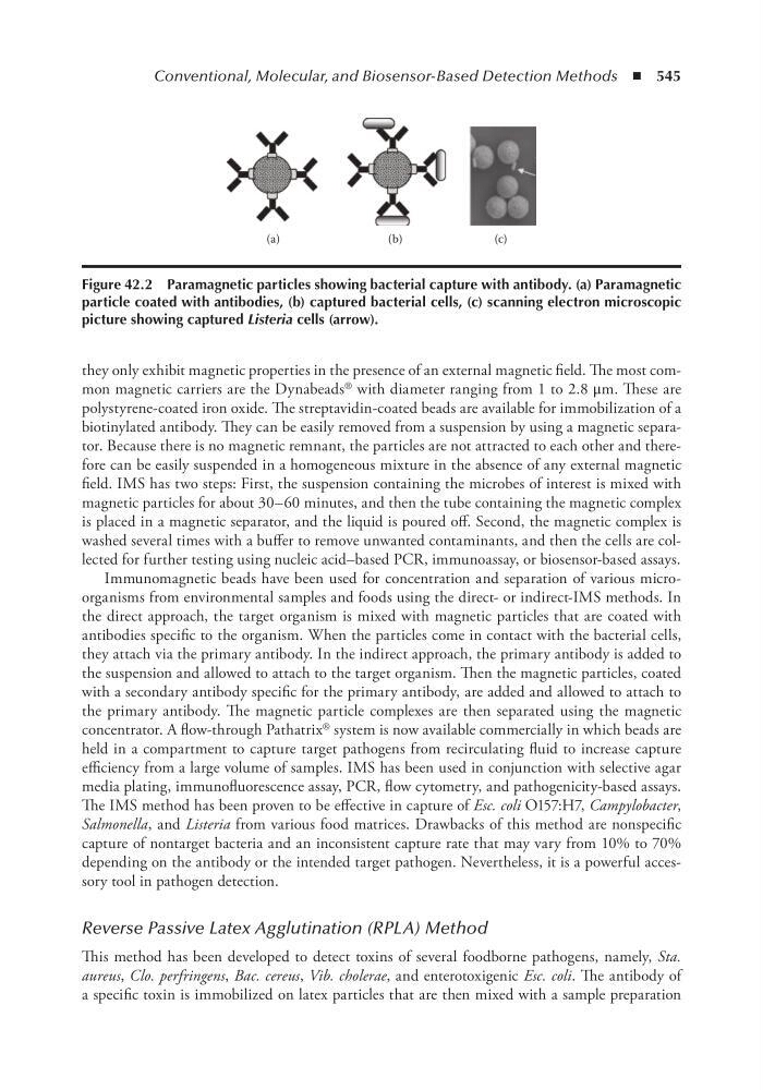
Figure 42.2 Paramagnetic particles showing bacterial capture with antibody. (a) Paramagnetic
particle coated with antibodies, (b) captured bacterial cells, (c) scanning electron microscopic
picture showing captured Listeria cells (arrow).
they only exhibit magnetic properties in the presence of an external magnetic field. The most common magnetic carriers are the Dynabeads ® with diameter ranging from 1 to 2.8 μm. These are
polystyrene-coated iron oxide. The streptavidin-coated beads are available for immobilization of a
biotinylated antibody. They can be easily removed from a suspension by using a magnetic separator. Because there is no magnetic remnant, the particles are not attracted to each other and therefore can be easily suspended in a homogeneous mixture in the absence of any external magnetic
field. IMS has two steps: First, the suspension containing the microbes of interest is mixed with
magnetic particles for about 30–60 minutes, and then the tube containing the magnetic complex
is placed in a magnetic separator, and the liquid is poured off. Second, the magnetic complex is
washed several times with a buffer to remove unwanted contaminants, and then the cells are collected for further testing using nucleic acid–based PCR, immunoassay, or biosensor-based assays.
Immunomagnetic beads have been used for concentration and separation of various microorganisms from environmental samples and foods using the direct- or indirect-IMS methods. In
the direct approach, the target organism is mixed with magnetic particles that are coated with
antibodies specific to the organism. When the particles come in contact with the bacterial cells,
they attach via the primary antibody. In the indirect approach, the primary antibody is added to
the suspension and allowed to attach to the target organism. Then the magnetic particles, coated
with a secondary antibody specific for the primary antibody, are added and allowed to attach to
the primary antibody. The magnetic particle complexes are then separated using the magnetic
concentrator. A flow-through Pathatrix ® system is now available commercially in which beads are
held in a compartment to capture target pathogens from recirculating fluid to increase capture
efficiency from a large volume of samples. IMS has been used in conjunction with selective agar
media plating, immunofluorescence assay, PCR, flow cytometry, and pathogenicity-based assays.
The IMS method has been proven to be effective in capture of Esc. coli O157:H7, Campylobacter,
Salmonella, and Listeria from various food matrices. Drawbacks of this method are nonspecific
capture of nontarget bacteria and an inconsistent capture rate that may vary from 10% to 70%
depending on the antibody or the intended target pathogen. Nevertheless, it is a powerful accessory tool in pathogen detection.
Reverse Passive Latex Agglutination (RPLA) Method
This method has been developed to detect toxins of several foodborne pathogens, namely, Sta.
aureus, Clo. perfringens, Bac. cereus, Vib. cholerae, and enterotoxigenic Esc. coli. The antibody of
a specific toxin is immobilized on latex particles that are then mixed with a sample preparation
(a)
(b)
(c)
Figure 42.2 Paramagnetic particles showing bacterial capture with antibody. (a) Paramagnetic
particle coated with antibodies, (b) captured bacterial cells, (c) scanning electron microscopic
picture showing captured Listeria cells (arrow).