

Foodborne Bacterial Infections ◾ 347
of 10 8 –10 10 cells. Most often, the symptoms appear within one to seven days following ingestion
and include mild flu-like symptoms with slight fever, abdominal cramps, and diarrhea. The symptoms subside in a few days, but the individual sheds Lis. monocytogenes in the feces for some time.
Invasive Systemic Disease 13–15
This form of disease is associated with immunologically challenged populations. These groups
include pregnant women, unborn fetuses, infants, elderly people with reduced immunity from diseases, and people taking special medications, such as steroids and chemotherapeutic agents to treat
cancer. The infective dose in these people is considered to be about 100–1000 cells. Following
ingestion, bacteria pass through the stomach unharmed and then reach the small intestine. GAD
(glutamate decarboxylase) helps bacterial survival in the stomach acid while bile salt hydrolase
protects Lis. monocytogenes from antimicrobial action of bile salts. Bacteria rapidly pass through
the intestine via the Peyer’s patch, a localized lymphatic tissue, where M cells phagocytose and
allow bacterial translocation to the lamina propria and then to blood circulation. Lis. monocytogenes can also actively pass through the epithelial barrier (Figure 26.2). First, bacteria bind to
epithelial cells using adhesion proteins like Listeria adhesion protein (LAP), fibronectin binding
protein (Fbp), and amidase and then invade epithelial cells using Internalin A, Internalin B, LapB,
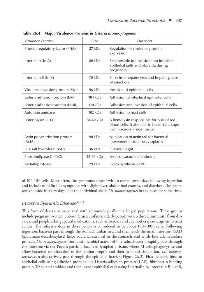
table 26.4 Major Virulence Proteins in Listeria monocytogenes
Virulence Factors
Size
Function
Protein regulatory factor (PrfA)
27 kDa
Regulation of virulence protein
expression
Internalin (InlA)
80 kDa
Responsible for invasion into intestinal
epithelial cells and placenta during
pregnancy
Internalin B (InlB)
70 kDa
Entry into hepatocytes and hepatic phase
of infection
Virulence invasion protein (Vip)
96 kDa
Invasion of epithelial cells
Listeria adhesion protein (LAP)
104 kDa
Adhesion to intestinal epithelial cells
Listeria adhesion protein (LapB)
170 kDa
Adhesion and invasion of epithelial cells
Autolysin amidase
102 kDa
Adhesion to host cells
Listeriolysin (LLO)
58–60 kDa
A hemolysin responsible for lysis of red
blood cells. It also aids in bacterial escape
from vacuole inside the cell
Actin polymerization protein
(ActA)
90 kDa
Nucleation of actin tail for bacterial
movement inside the cytoplasm
Bile salt hydrolase (BSH)
36 kDa
Survival in gut
Phospholipase C (PLC)
29–33 kDa
Lyses of vacuole membrane
Metalloprotease
29 kDa
Helps synthesis of PLC
of 10 8 –10 10 cells. Most often, the symptoms appear within one to seven days following ingestion
and include mild flu-like symptoms with slight fever, abdominal cramps, and diarrhea. The symptoms subside in a few days, but the individual sheds Lis. monocytogenes in the feces for some time.
Invasive Systemic Disease 13–15
This form of disease is associated with immunologically challenged populations. These groups
include pregnant women, unborn fetuses, infants, elderly people with reduced immunity from diseases, and people taking special medications, such as steroids and chemotherapeutic agents to treat
cancer. The infective dose in these people is considered to be about 100–1000 cells. Following
ingestion, bacteria pass through the stomach unharmed and then reach the small intestine. GAD
(glutamate decarboxylase) helps bacterial survival in the stomach acid while bile salt hydrolase
protects Lis. monocytogenes from antimicrobial action of bile salts. Bacteria rapidly pass through
the intestine via the Peyer’s patch, a localized lymphatic tissue, where M cells phagocytose and
allow bacterial translocation to the lamina propria and then to blood circulation. Lis. monocytogenes can also actively pass through the epithelial barrier (Figure 26.2). First, bacteria bind to
epithelial cells using adhesion proteins like Listeria adhesion protein (LAP), fibronectin binding
protein (Fbp), and amidase and then invade epithelial cells using Internalin A, Internalin B, LapB,
table 26.4 Major Virulence Proteins in Listeria monocytogenes
Virulence Factors
Size
Function
Protein regulatory factor (PrfA)
27 kDa
Regulation of virulence protein
expression
Internalin (InlA)
80 kDa
Responsible for invasion into intestinal
epithelial cells and placenta during
pregnancy
Internalin B (InlB)
70 kDa
Entry into hepatocytes and hepatic phase
of infection
Virulence invasion protein (Vip)
96 kDa
Invasion of epithelial cells
Listeria adhesion protein (LAP)
104 kDa
Adhesion to intestinal epithelial cells
Listeria adhesion protein (LapB)
170 kDa
Adhesion and invasion of epithelial cells
Autolysin amidase
102 kDa
Adhesion to host cells
Listeriolysin (LLO)
58–60 kDa
A hemolysin responsible for lysis of red
blood cells. It also aids in bacterial escape
from vacuole inside the cell
Actin polymerization protein
(ActA)
90 kDa
Nucleation of actin tail for bacterial
movement inside the cytoplasm
Bile salt hydrolase (BSH)
36 kDa
Survival in gut
Phospholipase C (PLC)
29–33 kDa
Lyses of vacuole membrane
Metalloprotease
29 kDa
Helps synthesis of PLC