

101
used to determine the molar ratio of tri- to tetrasaccharide (DP3:DP4). This molar
ratio varies, but is typically about 2.1 for oats, 3.2 for barley and 3.5 for wheat
(Wood et al. 1991a, b), and the ratio always is higher for barley than oats (Cui et al.
2000; Tosh et al. 2004). HPAEC-assisted lichenase treatment can be used to determine barley β-glucan structure and molar ratio of 1→4 to 1→3 linkages for barley
varieties ranged from 2.26 to 2.35 (Yoo et al. 2007). High-performance sizeexclusion chromatography (HPSEC) can be used to determine the molar mass of
β-glucans. Molecular weight is important in influencing β-glucan solubility as
500,000 g/mol molecular weight has been demonstrated for soluble β-glucans of
both oat and barley and <200,000 g/mol for insoluble β-glucans (Johansson et al.
2004). Molecular weight of β-glucans varies greatly with oats (0.065–3 × 10⁶) typically higher than barley (0.15–2.5 × 10⁶) or wheat (0.25–0.7 × 10⁶) (Andersson
et al. 2004; Beer et al. 1997; Lazaridou et al. 2003).
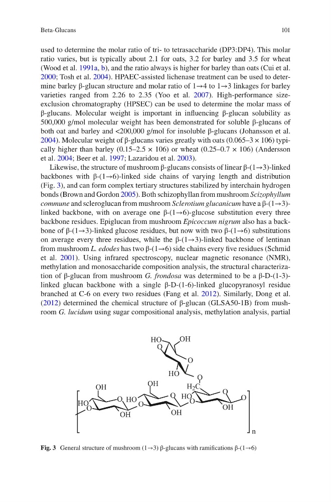
Likewise, the structure of mushroom β-glucans consists of linear β-(1→3)-linked
backbones with β-(1→6)-linked side chains of varying length and distribution
(Fig. 3), and can form complex tertiary structures stabilized by interchain hydrogen
bonds (Brown and Gordon 2005). Both schizophyllan from mushroom Scizophyllum
commune and scleroglucan from mushroom Sclerotium glucanicum have a β-(1→3)linked backbone, with on average one β-(1→6)-glucose substitution every three
backbone residues. Epiglucan from mushroom Epicoccum nigrum also has a backbone of β-(1→3)-linked glucose residues, but now with two β-(1→6) substitutions
on average every three residues, while the β-(1→3)-linked backbone of lentinan
from mushroom L. edodes has two β-(1→6) side chains every five residues (Schmid
et al. 2001). Using infrared spectroscopy, nuclear magnetic resonance (NMR),
methylation and monosaccharide composition analysis, the structural characterization of β-glucan from mushroom G. frondosa was determined to be a β-D-(1-3)linked glucan backbone with a single β-D-(1-6)-linked glucopyranosyl residue
branched at C-6 on every two residues (Fang et al. 2012). Similarly, Dong et al.
(2012) determined the chemical structure of β-glucan (GLSA50-1B) from mushroom G. lucidum using sugar compositional analysis, methylation analysis, partial
OH
OH
H 2 C
n
OH
HO
HO
HO
HO
OH
OH
OH
HO
O
O
O
O
O
O
O
O
O
O
Fig. 3 General structure of mushroom (1→3) β-glucans with ramifications β-(1→6)
Beta-Glucans
used to determine the molar ratio of tri- to tetrasaccharide (DP3:DP4). This molar
ratio varies, but is typically about 2.1 for oats, 3.2 for barley and 3.5 for wheat
(Wood et al. 1991a, b), and the ratio always is higher for barley than oats (Cui et al.
2000; Tosh et al. 2004). HPAEC-assisted lichenase treatment can be used to determine barley β-glucan structure and molar ratio of 1→4 to 1→3 linkages for barley
varieties ranged from 2.26 to 2.35 (Yoo et al. 2007). High-performance sizeexclusion chromatography (HPSEC) can be used to determine the molar mass of
β-glucans. Molecular weight is important in influencing β-glucan solubility as
500,000 g/mol molecular weight has been demonstrated for soluble β-glucans of
both oat and barley and <200,000 g/mol for insoluble β-glucans (Johansson et al.
2004). Molecular weight of β-glucans varies greatly with oats (0.065–3 × 10⁶) typically higher than barley (0.15–2.5 × 10⁶) or wheat (0.25–0.7 × 10⁶) (Andersson
et al. 2004; Beer et al. 1997; Lazaridou et al. 2003).
Likewise, the structure of mushroom β-glucans consists of linear β-(1→3)-linked
backbones with β-(1→6)-linked side chains of varying length and distribution
(Fig. 3), and can form complex tertiary structures stabilized by interchain hydrogen
bonds (Brown and Gordon 2005). Both schizophyllan from mushroom Scizophyllum
commune and scleroglucan from mushroom Sclerotium glucanicum have a β-(1→3)linked backbone, with on average one β-(1→6)-glucose substitution every three
backbone residues. Epiglucan from mushroom Epicoccum nigrum also has a backbone of β-(1→3)-linked glucose residues, but now with two β-(1→6) substitutions
on average every three residues, while the β-(1→3)-linked backbone of lentinan
from mushroom L. edodes has two β-(1→6) side chains every five residues (Schmid
et al. 2001). Using infrared spectroscopy, nuclear magnetic resonance (NMR),
methylation and monosaccharide composition analysis, the structural characterization of β-glucan from mushroom G. frondosa was determined to be a β-D-(1-3)linked glucan backbone with a single β-D-(1-6)-linked glucopyranosyl residue
branched at C-6 on every two residues (Fang et al. 2012). Similarly, Dong et al.
(2012) determined the chemical structure of β-glucan (GLSA50-1B) from mushroom G. lucidum using sugar compositional analysis, methylation analysis, partial
OH
OH
H 2 C
n
OH
HO
HO
HO
HO
OH
OH
OH
HO
O
O
O
O
O
O
O
O
O
O
Fig. 3 General structure of mushroom (1→3) β-glucans with ramifications β-(1→6)
Beta-Glucans