

100
charide (Yang and Zhang 2009). Polysaccharide with a broad polydispersity can be
fractionated by stepwise precipitation or preparative gel permeation chromatography, through which polysaccharides with different molecular weights and low polydispersity can be achieved (Zhang et al. 2007).
Structural and Solution Behavior
Structural Features
Polysaccharides derived from different sources are usually composed of various
monosaccharides, and many of them have hyperbranched structures. To determine
the structure of glucans from different resources, a series of analytical methods have
been developed. The chemical structure was analyzed by Fourier transform infrared
spectroscopy (FITR), nuclear magnetic resonance spectroscopy (NMR), gas chromatography (GC), gas chromatography-mass spectrometer (GC-MS), and highperformance liquid chromatography (HPLC).
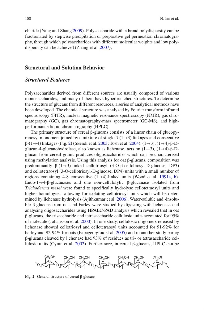
The primary structure of cereal β-glucans consists of a linear chain of glucopyranosyl monomers joined by a mixture of single β-(1→3) linkages and consecutive
β-(1→4) linkages (Fig. 2) (Skendi et al. 2003; Tosh et al. 2004). (1→3), (1→4)-β-Dglucan-4-glucanohydrolase, also known as lichenase, acts on (1→3), (1→4)-β-Dglucan from cereal grains produces oligosaccharides which can be characterised
using methylation analysis. Using this analysis for oat β-glucans, composition was
predominantly β-(1→3)-linked cellotriosyl (3-O-β-cellobiosyl-D-glucose, DP3)
and cellotetraosyl (3-O-cellotriosyl-D-glucose, DP4) units with a small number of
regions containing 4-8 consecutive (1→4)-linked units (Wood et al. 1991a, b).
Endo-1→4-β-glucanases and one non-cellulolytic β-glucanase isolated from
Trichoderma reesei were found to specifically hydrolyse cellotetraosyl units and
higher homologues, allowing for isolating cellotriosyl units which will be determined by lichenase hydrolysis (Ajithkumar et al. 2006). Water-soluble and -insoluble β-glucans from oat and barley were studied by digesting with lichenase and
analysing oligosaccharides using HPAEC-PAD analysis which revealed that in oat
β-glucans, the trisaccharide and tetrasaccharide cellulosic units accounted for 95%
of molecule (Johansson et al. 2000). In one study, cellulosic oligomers released by
lichenase showed cellotriosyl and cellotetraosyl units accounted for 91-92% for
barley and 92-94% for oats (Papageorgiou et al. 2005) and in another study barley
β-glucans cleaved by lichenase had 93% of residues as tri- or tetrasaccharide cellulosic units (Cyran et al. 2002). Furthermore, in cereal β-glucans, HPLC can be
O
CH 2 OH
CH 2 OH CH 2 OH
CH 2 OH
CH 2 OH CH 2 OH
CH 2 OH
O
O
O
O O
O
O
O
O
O
O
O
O
O
Fig. 2 General structure of cereal β-glucans
N. Jan et al.
charide (Yang and Zhang 2009). Polysaccharide with a broad polydispersity can be
fractionated by stepwise precipitation or preparative gel permeation chromatography, through which polysaccharides with different molecular weights and low polydispersity can be achieved (Zhang et al. 2007).
Structural and Solution Behavior
Structural Features
Polysaccharides derived from different sources are usually composed of various
monosaccharides, and many of them have hyperbranched structures. To determine
the structure of glucans from different resources, a series of analytical methods have
been developed. The chemical structure was analyzed by Fourier transform infrared
spectroscopy (FITR), nuclear magnetic resonance spectroscopy (NMR), gas chromatography (GC), gas chromatography-mass spectrometer (GC-MS), and highperformance liquid chromatography (HPLC).
The primary structure of cereal β-glucans consists of a linear chain of glucopyranosyl monomers joined by a mixture of single β-(1→3) linkages and consecutive
β-(1→4) linkages (Fig. 2) (Skendi et al. 2003; Tosh et al. 2004). (1→3), (1→4)-β-Dglucan-4-glucanohydrolase, also known as lichenase, acts on (1→3), (1→4)-β-Dglucan from cereal grains produces oligosaccharides which can be characterised
using methylation analysis. Using this analysis for oat β-glucans, composition was
predominantly β-(1→3)-linked cellotriosyl (3-O-β-cellobiosyl-D-glucose, DP3)
and cellotetraosyl (3-O-cellotriosyl-D-glucose, DP4) units with a small number of
regions containing 4-8 consecutive (1→4)-linked units (Wood et al. 1991a, b).
Endo-1→4-β-glucanases and one non-cellulolytic β-glucanase isolated from
Trichoderma reesei were found to specifically hydrolyse cellotetraosyl units and
higher homologues, allowing for isolating cellotriosyl units which will be determined by lichenase hydrolysis (Ajithkumar et al. 2006). Water-soluble and -insoluble β-glucans from oat and barley were studied by digesting with lichenase and
analysing oligosaccharides using HPAEC-PAD analysis which revealed that in oat
β-glucans, the trisaccharide and tetrasaccharide cellulosic units accounted for 95%
of molecule (Johansson et al. 2000). In one study, cellulosic oligomers released by
lichenase showed cellotriosyl and cellotetraosyl units accounted for 91-92% for
barley and 92-94% for oats (Papageorgiou et al. 2005) and in another study barley
β-glucans cleaved by lichenase had 93% of residues as tri- or tetrasaccharide cellulosic units (Cyran et al. 2002). Furthermore, in cereal β-glucans, HPLC can be
O
CH 2 OH
CH 2 OH CH 2 OH
CH 2 OH
CH 2 OH CH 2 OH
CH 2 OH
O
O
O
O O
O
O
O
O
O
O
O
O
O
Fig. 2 General structure of cereal β-glucans
N. Jan et al.