

From Plant to Patient: Thapsigargin, a Tool for Understanding …
85
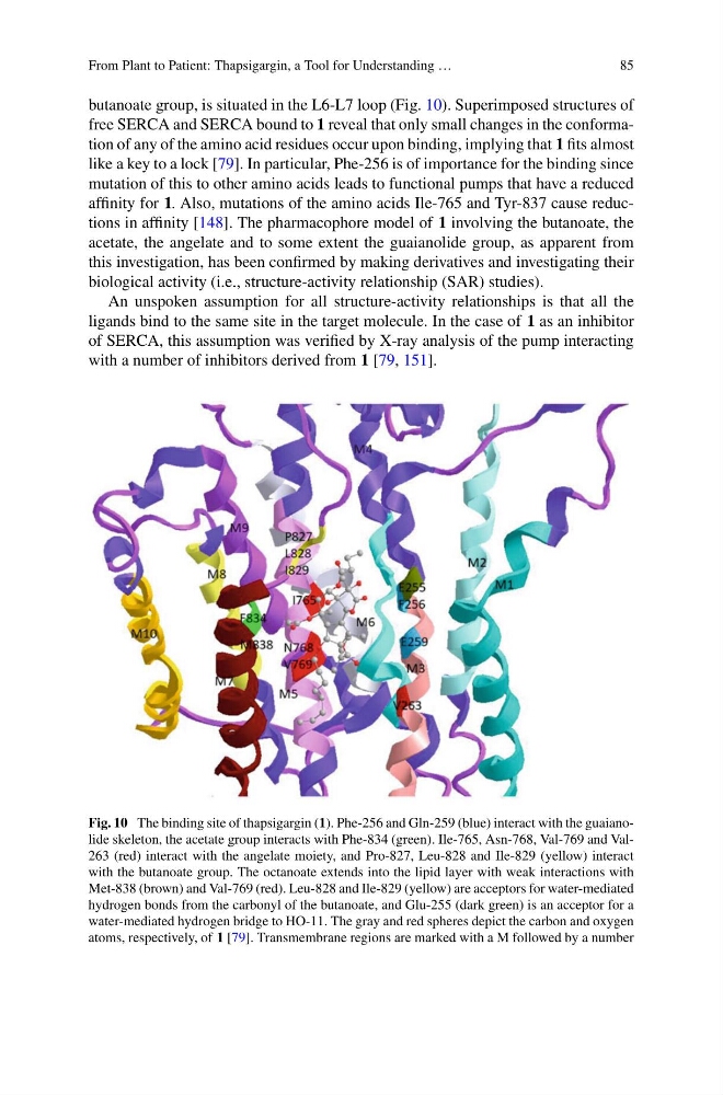
butanoate group, is situated in the L6-L7 loop (Fig. 10). Superimposed structures of
free SERCA and SERCA bound to 1 reveal that only small changes in the conformation of any of the amino acid residues occur upon binding, implying that 1 fits almost
like a key to a lock [79]. In particular, Phe-256 is of importance for the binding since
mutation of this to other amino acids leads to functional pumps that have a reduced
affinity for 1. Also, mutations of the amino acids Ile-765 and Tyr-837 cause reductions in affinity [148]. The pharmacophore model of 1 involving the butanoate, the
acetate, the angelate and to some extent the guaianolide group, as apparent from
this investigation, has been confirmed by making derivatives and investigating their
biological activity (i.e., structure-activity relationship (SAR) studies).
An unspoken assumption for all structure-activity relationships is that all the
ligands bind to the same site in the target molecule. In the case of 1 as an inhibitor
of SERCA, this assumption was verified by X-ray analysis of the pump interacting
with a number of inhibitors derived from 1 [79, 151].
Fig. 10 The binding site of thapsigargin (1). Phe-256 and Gln-259 (blue) interact with the guaianolide skeleton, the acetate group interacts with Phe-834 (green). Ile-765, Asn-768, Val-769 and Val263 (red) interact with the angelate moiety, and Pro-827, Leu-828 and Ile-829 (yellow) interact
with the butanoate group. The octanoate extends into the lipid layer with weak interactions with
Met-838 (brown) and Val-769 (red). Leu-828 and Ile-829 (yellow) are acceptors for water-mediated
hydrogen bonds from the carbonyl of the butanoate, and Glu-255 (dark green) is an acceptor for a
water-mediated hydrogen bridge to HO-11. The gray and red spheres depict the carbon and oxygen
atoms, respectively, of 1 [79]. Transmembrane regions are marked with a M followed by a number
85
butanoate group, is situated in the L6-L7 loop (Fig. 10). Superimposed structures of
free SERCA and SERCA bound to 1 reveal that only small changes in the conformation of any of the amino acid residues occur upon binding, implying that 1 fits almost
like a key to a lock [79]. In particular, Phe-256 is of importance for the binding since
mutation of this to other amino acids leads to functional pumps that have a reduced
affinity for 1. Also, mutations of the amino acids Ile-765 and Tyr-837 cause reductions in affinity [148]. The pharmacophore model of 1 involving the butanoate, the
acetate, the angelate and to some extent the guaianolide group, as apparent from
this investigation, has been confirmed by making derivatives and investigating their
biological activity (i.e., structure-activity relationship (SAR) studies).
An unspoken assumption for all structure-activity relationships is that all the
ligands bind to the same site in the target molecule. In the case of 1 as an inhibitor
of SERCA, this assumption was verified by X-ray analysis of the pump interacting
with a number of inhibitors derived from 1 [79, 151].
Fig. 10 The binding site of thapsigargin (1). Phe-256 and Gln-259 (blue) interact with the guaianolide skeleton, the acetate group interacts with Phe-834 (green). Ile-765, Asn-768, Val-769 and Val263 (red) interact with the angelate moiety, and Pro-827, Leu-828 and Ile-829 (yellow) interact
with the butanoate group. The octanoate extends into the lipid layer with weak interactions with
Met-838 (brown) and Val-769 (red). Leu-828 and Ile-829 (yellow) are acceptors for water-mediated
hydrogen bonds from the carbonyl of the butanoate, and Glu-255 (dark green) is an acceptor for a
water-mediated hydrogen bridge to HO-11. The gray and red spheres depict the carbon and oxygen
atoms, respectively, of 1 [79]. Transmembrane regions are marked with a M followed by a number