

80
S. B. Christensen et al.
O
O
O
O
OH
OH
O
O
O
O
O
O
O
OH
OH
O
O
O
a
O
O
O
O
O
OH
OH
O
O
O
b
O
O
6
O
O
O
O
OH
OH
O
HO
O
c
O
O
6
O
O
O
O
OH
OH
O
O
O
i
O
O
6
O
d
17
1
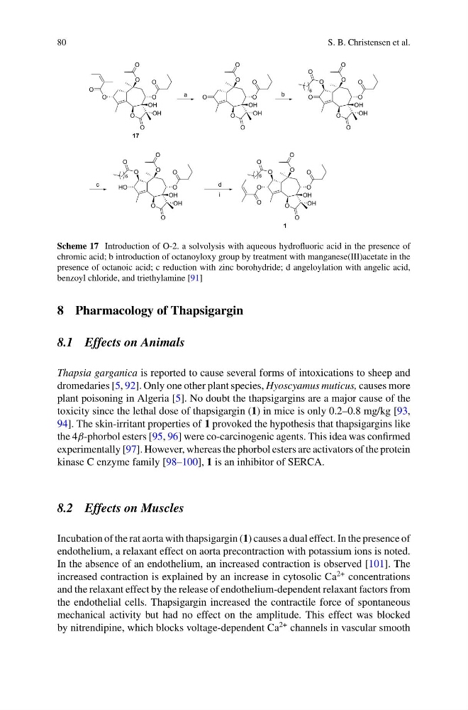
Scheme 17 Introduction of O-2. a solvolysis with aqueous hydrofluoric acid in the presence of
chromic acid; b introduction of octanoyloxy group by treatment with manganese(III)acetate in the
presence of octanoic acid; c reduction with zinc borohydride; d angeloylation with angelic acid,
benzoyl chloride, and triethylamine [91]
8 Pharmacology of Thapsigargin
8.1 Effects on Animals
Thapsia garganica is reported to cause several forms of intoxications to sheep and
dromedaries [5, 92]. Only one other plant species, Hyoscyamus muticus, causes more
plant poisoning in Algeria [5]. No doubt the thapsigargins are a major cause of the
toxicity since the lethal dose of thapsigargin (1) in mice is only 0.2–0.8 mg/kg [93,
94]. The skin-irritant properties of 1 provoked the hypothesis that thapsigargins like
the 4β-phorbol esters [95, 96] were co-carcinogenic agents. This idea was confirmed
experimentally [97]. However, whereas the phorbol esters are activators of the protein
kinase C enzyme family [98–100], 1 is an inhibitor of SERCA.
8.2 Effects on Muscles
Incubation of the rat aorta with thapsigargin (1) causes a dual effect. In the presence of
endothelium, a relaxant effect on aorta precontraction with potassium ions is noted.
In the absence of an endothelium, an increased contraction is observed [101]. The
increased contraction is explained by an increase in cytosolic Ca
2+ concentrations
and the relaxant effect by the release of endothelium-dependent relaxant factors from
the endothelial cells. Thapsigargin increased the contractile force of spontaneous
mechanical activity but had no effect on the amplitude. This effect was blocked
by nitrendipine, which blocks voltage-dependent Ca
2+ channels in vascular smooth
S. B. Christensen et al.
O
O
O
O
OH
OH
O
O
O
O
O
O
O
OH
OH
O
O
O
a
O
O
O
O
O
OH
OH
O
O
O
b
O
O
6
O
O
O
O
OH
OH
O
HO
O
c
O
O
6
O
O
O
O
OH
OH
O
O
O
i
O
O
6
O
d
17
1
Scheme 17 Introduction of O-2. a solvolysis with aqueous hydrofluoric acid in the presence of
chromic acid; b introduction of octanoyloxy group by treatment with manganese(III)acetate in the
presence of octanoic acid; c reduction with zinc borohydride; d angeloylation with angelic acid,
benzoyl chloride, and triethylamine [91]
8 Pharmacology of Thapsigargin
8.1 Effects on Animals
Thapsia garganica is reported to cause several forms of intoxications to sheep and
dromedaries [5, 92]. Only one other plant species, Hyoscyamus muticus, causes more
plant poisoning in Algeria [5]. No doubt the thapsigargins are a major cause of the
toxicity since the lethal dose of thapsigargin (1) in mice is only 0.2–0.8 mg/kg [93,
94]. The skin-irritant properties of 1 provoked the hypothesis that thapsigargins like
the 4β-phorbol esters [95, 96] were co-carcinogenic agents. This idea was confirmed
experimentally [97]. However, whereas the phorbol esters are activators of the protein
kinase C enzyme family [98–100], 1 is an inhibitor of SERCA.
8.2 Effects on Muscles
Incubation of the rat aorta with thapsigargin (1) causes a dual effect. In the presence of
endothelium, a relaxant effect on aorta precontraction with potassium ions is noted.
In the absence of an endothelium, an increased contraction is observed [101]. The
increased contraction is explained by an increase in cytosolic Ca
2+ concentrations
and the relaxant effect by the release of endothelium-dependent relaxant factors from
the endothelial cells. Thapsigargin increased the contractile force of spontaneous
mechanical activity but had no effect on the amplitude. This effect was blocked
by nitrendipine, which blocks voltage-dependent Ca
2+ channels in vascular smooth