

9.6 Luminescent Nanocomposites 211
molecules bind, similarly as m-PMMA, to the surface forming H–(C=O)–O–
(oxide particle).
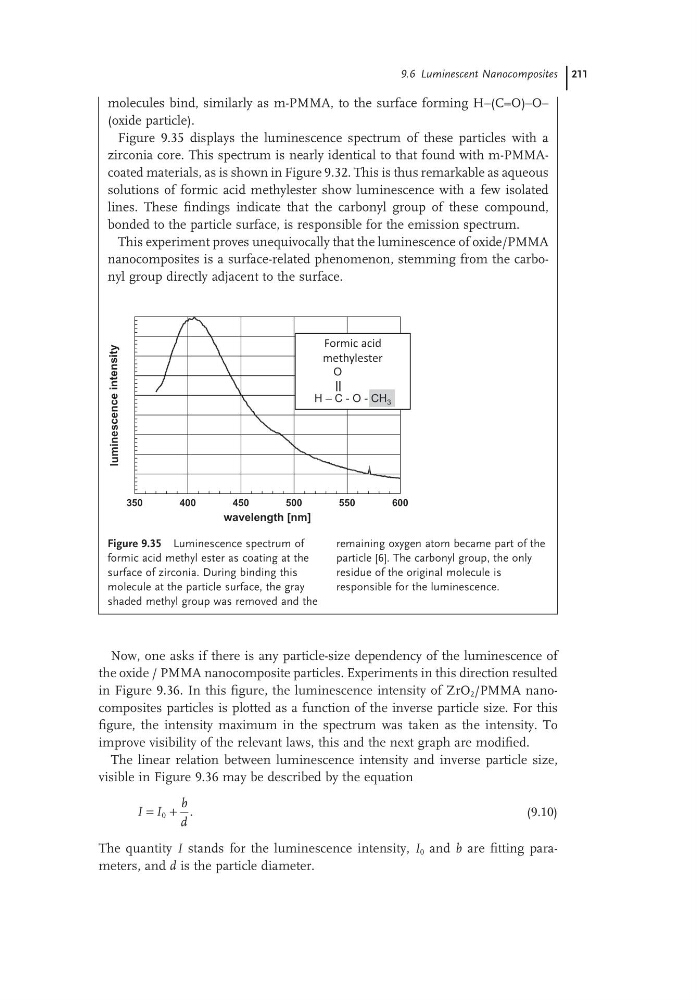
Figure 9.35 displays the luminescence spectrum of these particles with a
zirconia core. This spectrum is nearly identical to that found with m-PMMAcoated materials, as is shown in Figure 9.32. This is thus remarkable as aqueous
solutions of formic acid methylester show luminescence with a few isolated
lines. These findings indicate that the carbonyl group of these compound,
bonded to the particle surface, is responsible for the emission spectrum.
This experiment proves unequivocally that the luminescence of oxide/PMMA
nanocomposites is a surface-related phenomenon, stemming from the carbonyl group directly adjacent to the surface.
Figure 9.35 Luminescence spectrum of
formic acid methyl ester as coating at the
surface of zirconia. During binding this
molecule at the particle surface, the gray
shaded methyl group was removed and the
remaining oxygen atom became part of the
particle [6]. The carbonyl group, the only
residue of the original molecule is
responsible for the luminescence.
350
400
450
500
550
600
wavelength [nm]
2
3
4
5
6
7
8
9
10
11
luminescence
intensity
Formic acid
methylester
O
||
H – C - O - CH 3
Now, one asks if there is any particle-size dependency of the luminescence of
the oxide / PMMA nanocomposite particles. Experiments in this direction resulted
in Figure 9.36. In this figure, the luminescence intensity of ZrO 2 /PMMA nanocomposites particles is plotted as a function of the inverse particle size. For this
figure, the intensity maximum in the spectrum was taken as the intensity. To
improve visibility of the relevant laws, this and the next graph are modified.
The linear relation between luminescence intensity and inverse particle size,
visible in Figure 9.36 may be described by the equation
I I
b
d
= +
0
.
(9.10)
The quantity I stands for the luminescence intensity, I 0 and b are fitting parameters, and d is the particle diameter.
molecules bind, similarly as m-PMMA, to the surface forming H–(C=O)–O–
(oxide particle).
Figure 9.35 displays the luminescence spectrum of these particles with a
zirconia core. This spectrum is nearly identical to that found with m-PMMAcoated materials, as is shown in Figure 9.32. This is thus remarkable as aqueous
solutions of formic acid methylester show luminescence with a few isolated
lines. These findings indicate that the carbonyl group of these compound,
bonded to the particle surface, is responsible for the emission spectrum.
This experiment proves unequivocally that the luminescence of oxide/PMMA
nanocomposites is a surface-related phenomenon, stemming from the carbonyl group directly adjacent to the surface.
Figure 9.35 Luminescence spectrum of
formic acid methyl ester as coating at the
surface of zirconia. During binding this
molecule at the particle surface, the gray
shaded methyl group was removed and the
remaining oxygen atom became part of the
particle [6]. The carbonyl group, the only
residue of the original molecule is
responsible for the luminescence.
350
400
450
500
550
600
wavelength [nm]
2
3
4
5
6
7
8
9
10
11
luminescence
intensity
Formic acid
methylester
O
||
H – C - O - CH 3
Now, one asks if there is any particle-size dependency of the luminescence of
the oxide / PMMA nanocomposite particles. Experiments in this direction resulted
in Figure 9.36. In this figure, the luminescence intensity of ZrO 2 /PMMA nanocomposites particles is plotted as a function of the inverse particle size. For this
figure, the intensity maximum in the spectrum was taken as the intensity. To
improve visibility of the relevant laws, this and the next graph are modified.
The linear relation between luminescence intensity and inverse particle size,
visible in Figure 9.36 may be described by the equation
I I
b
d
= +
0
.
(9.10)
The quantity I stands for the luminescence intensity, I 0 and b are fitting parameters, and d is the particle diameter.