

2. Laboratory glass bottles with caps (25 mL, Duran) to store
biomaterial powder.
3. Glass beaker (20 or 40 mL volume capacity, depending on the
amount of biopaste needed).
4. Magnetic stirrer bar.
5. Laponite XLG (BYK Additives & Instruments).
6. Alginate (alginic acid sodium salt from brown algae, mannuronic/glucuronic (M/G) acid ratio 1:2).
7. Methylcellulose (MW % 88 kDa, 4000 cP).
8. Sterile stainless steel spatulas.
9. Magnetic stirrer (reaching 28 RCF to 300 RPM).
2.2 Cell Culture
1. Skeletal stem cells (SSCs; isolated from human sources with full
national ethical approval and after patient consent acquired).
2. Cell culture flasks (175 cm
2 cell culture flask, angled neck,
non-pyrogenic polystyrene).
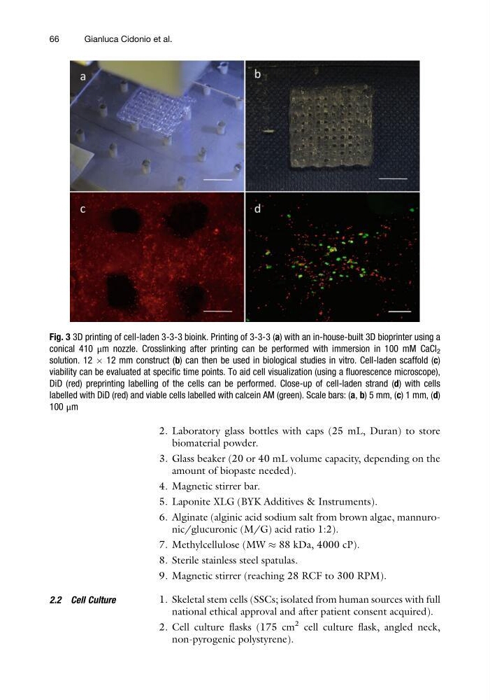
Fig. 3 3D printing of cell-laden 3-3-3 bioink. Printing of 3-3-3 (a) with an in-house-built 3D bioprinter using a
conical 410 μm nozzle. Crosslinking after printing can be performed with immersion in 100 mM CaCl 2
solution. 12 Â 12 mm construct (b) can then be used in biological studies in vitro. Cell-laden scaffold (c)
viability can be evaluated at specific time points. To aid cell visualization (using a fluorescence microscope),
DiD (red) preprinting labelling of the cells can be performed. Close-up of cell-laden strand (d) with cells
labelled with DiD (red) and viable cells labelled with calcein AM (green). Scale bars: (a, b) 5 mm, (c) 1 mm, (d)
100 μm
66
Gianluca Cidonio et al.
biomaterial powder.
3. Glass beaker (20 or 40 mL volume capacity, depending on the
amount of biopaste needed).
4. Magnetic stirrer bar.
5. Laponite XLG (BYK Additives & Instruments).
6. Alginate (alginic acid sodium salt from brown algae, mannuronic/glucuronic (M/G) acid ratio 1:2).
7. Methylcellulose (MW % 88 kDa, 4000 cP).
8. Sterile stainless steel spatulas.
9. Magnetic stirrer (reaching 28 RCF to 300 RPM).
2.2 Cell Culture
1. Skeletal stem cells (SSCs; isolated from human sources with full
national ethical approval and after patient consent acquired).
2. Cell culture flasks (175 cm
2 cell culture flask, angled neck,
non-pyrogenic polystyrene).
Fig. 3 3D printing of cell-laden 3-3-3 bioink. Printing of 3-3-3 (a) with an in-house-built 3D bioprinter using a
conical 410 μm nozzle. Crosslinking after printing can be performed with immersion in 100 mM CaCl 2
solution. 12 Â 12 mm construct (b) can then be used in biological studies in vitro. Cell-laden scaffold (c)
viability can be evaluated at specific time points. To aid cell visualization (using a fluorescence microscope),
DiD (red) preprinting labelling of the cells can be performed. Close-up of cell-laden strand (d) with cells
labelled with DiD (red) and viable cells labelled with calcein AM (green). Scale bars: (a, b) 5 mm, (c) 1 mm, (d)
100 μm
66
Gianluca Cidonio et al.