

4. CAD files can be converted to STL files by using Rhinoceros
software. The STL files can be sliced into 2D sections, each
having 25 μm layer thickness, through the EnvisionTEC Perfactory RP 2.0 software.
Acknowledgments
This work was partially supported by Short Term Mobility Project
2016 (STM 2016) “Photo-crosslinkable polymers for 3D-printing
of biomedical devices.”
References
1. Yan X, Gu P (1996) A review of rapid prototyping technologies and systems. Comput
Aided Des 28(4):307–318
2. Pham DT, Gault RS (1998) A comparison of
rapid prototyping technologies. Int J Mach
Tools Manuf 38(10–11):1257–1287
3. Kruth JP, Leu MC, Nakagawa T (1998) Progress in additive manufacturing and rapid prototyping. CIRP Ann 47(2):525–540
4. Eloma L, Kokkari A, N€ arhi T et al (2013)
Porous 3D modeled scaffolds of bioactive
glass
and
photocrosslinkable
poly
(e-caprolactone)
by
stereolithography.
Compos Sci Technol 74:99–106
5. Gloria A, Russo T, De Santis R et al (2009) 3D
fiber
deposition
technique
to
make
multifunctional and tailor-made scaffolds for
tissue engineering applications. J Appl Biomater Biomech 7:141–152
6. Rengier F et al (2010) 3D printing based on
imaging data: review of medical applications.
Int J Comput Assist Radiol Surg 5(4):335–341
7. Petrovic V et al (2011) Additive layered
manufacturing: sectors of industrial application
shown through case studies. Int J Prod Res 49
(4):1061–1079
8. Potamianos P et al (1998) Rapid prototyping
for orthopaedic surgery. Proc Inst Mech Eng H
J Eng Med 212(5):383–393
9. Puppi D, Chiellini F, Piras AM, Chiellini E
(2010) Polymeric materials for bone and cartilage repair. Prog Polym Sci 35(4):403–440
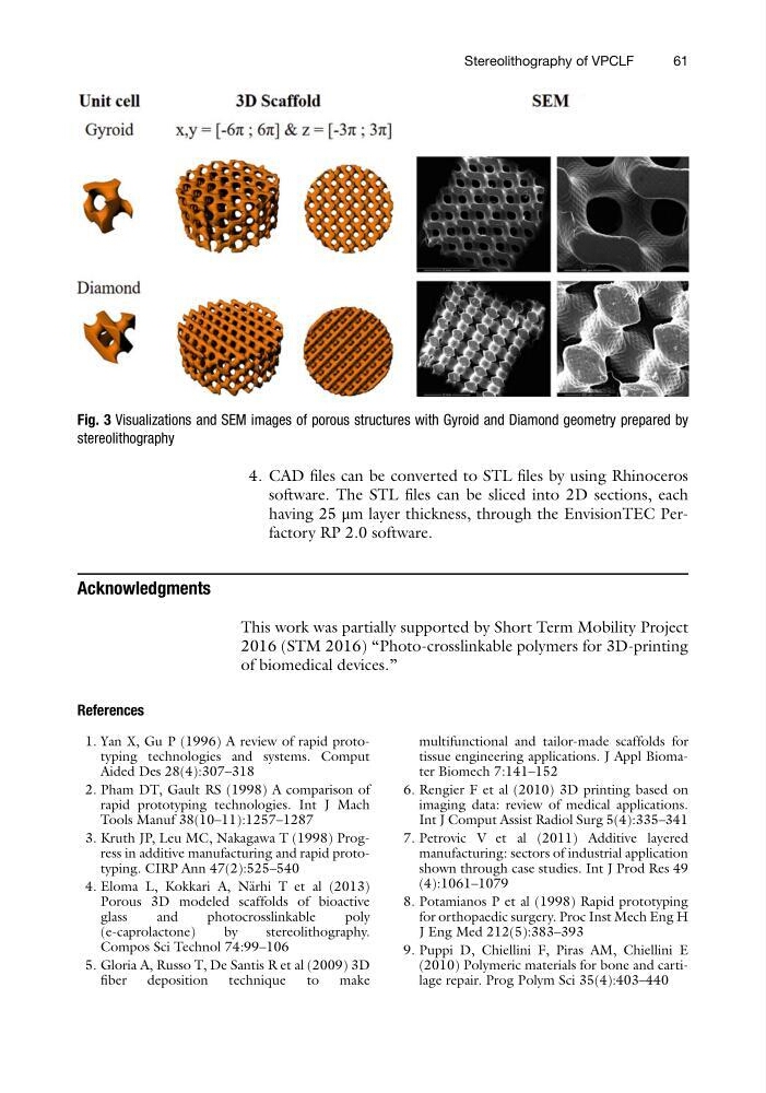
Fig. 3 Visualizations and SEM images of porous structures with Gyroid and Diamond geometry prepared by
stereolithography
Stereolithography of VPCLF
61
software. The STL files can be sliced into 2D sections, each
having 25 μm layer thickness, through the EnvisionTEC Perfactory RP 2.0 software.
Acknowledgments
This work was partially supported by Short Term Mobility Project
2016 (STM 2016) “Photo-crosslinkable polymers for 3D-printing
of biomedical devices.”
References
1. Yan X, Gu P (1996) A review of rapid prototyping technologies and systems. Comput
Aided Des 28(4):307–318
2. Pham DT, Gault RS (1998) A comparison of
rapid prototyping technologies. Int J Mach
Tools Manuf 38(10–11):1257–1287
3. Kruth JP, Leu MC, Nakagawa T (1998) Progress in additive manufacturing and rapid prototyping. CIRP Ann 47(2):525–540
4. Eloma L, Kokkari A, N€ arhi T et al (2013)
Porous 3D modeled scaffolds of bioactive
glass
and
photocrosslinkable
poly
(e-caprolactone)
by
stereolithography.
Compos Sci Technol 74:99–106
5. Gloria A, Russo T, De Santis R et al (2009) 3D
fiber
deposition
technique
to
make
multifunctional and tailor-made scaffolds for
tissue engineering applications. J Appl Biomater Biomech 7:141–152
6. Rengier F et al (2010) 3D printing based on
imaging data: review of medical applications.
Int J Comput Assist Radiol Surg 5(4):335–341
7. Petrovic V et al (2011) Additive layered
manufacturing: sectors of industrial application
shown through case studies. Int J Prod Res 49
(4):1061–1079
8. Potamianos P et al (1998) Rapid prototyping
for orthopaedic surgery. Proc Inst Mech Eng H
J Eng Med 212(5):383–393
9. Puppi D, Chiellini F, Piras AM, Chiellini E
(2010) Polymeric materials for bone and cartilage repair. Prog Polym Sci 35(4):403–440
Fig. 3 Visualizations and SEM images of porous structures with Gyroid and Diamond geometry prepared by
stereolithography
Stereolithography of VPCLF
61