

7. Metallic tip (inner diameters 0.3–0.5 mm).
8. Initiators: use glass slides (dimensions approximately
76 Â 26 Â 1 mm) for PEO-water and gelatin solutions. Cut
the silicon wafers (thickness 500–550 μm) into stripes with a
width of roughly 10 mm, which can be used for the PS-DMF
solutions.
9. Scotch tape.
10. Collecting substrates: a range of materials can be used, such as
glass coverslip (dimensions ~22 Â 22 Â 0.175 mm), silicon
wafer (dimensions ~20 Â 20 Â 0.5 mm), and PDMS film
(dimensions ~20 Â 20 Â 0.1 mm) prepared as described in [9].
11. Software control: a LabVIEW program was constructed to
control the x-y stage movement. Figure 3a shows an example
to create parallel linear patterns.
2.3 Gelatin
Crosslinking Reagents
1. 1-ethyl-3-(dimethyl-aminopropyl) carbodiimide hydrochloride (EDC).
2. N-hydroxyl succinimide (NHS).
3. Mix absolute ethanol with water at a volume ratio of 9:1.
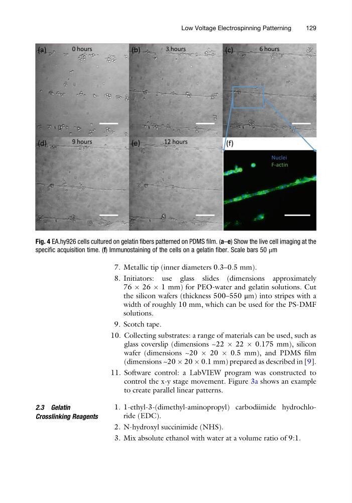
Fig. 4 EA.hy926 cells cultured on gelatin fibers patterned on PDMS film. (a–e) Show the live cell imaging at the
specific acquisition time. (f) Immunostaining of the cells on a gelatin fiber. Scale bars 50 μm
Low Voltage Electrospinning Patterning
129
8. Initiators: use glass slides (dimensions approximately
76 Â 26 Â 1 mm) for PEO-water and gelatin solutions. Cut
the silicon wafers (thickness 500–550 μm) into stripes with a
width of roughly 10 mm, which can be used for the PS-DMF
solutions.
9. Scotch tape.
10. Collecting substrates: a range of materials can be used, such as
glass coverslip (dimensions ~22 Â 22 Â 0.175 mm), silicon
wafer (dimensions ~20 Â 20 Â 0.5 mm), and PDMS film
(dimensions ~20 Â 20 Â 0.1 mm) prepared as described in [9].
11. Software control: a LabVIEW program was constructed to
control the x-y stage movement. Figure 3a shows an example
to create parallel linear patterns.
2.3 Gelatin
Crosslinking Reagents
1. 1-ethyl-3-(dimethyl-aminopropyl) carbodiimide hydrochloride (EDC).
2. N-hydroxyl succinimide (NHS).
3. Mix absolute ethanol with water at a volume ratio of 9:1.
Fig. 4 EA.hy926 cells cultured on gelatin fibers patterned on PDMS film. (a–e) Show the live cell imaging at the
specific acquisition time. (f) Immunostaining of the cells on a gelatin fiber. Scale bars 50 μm
Low Voltage Electrospinning Patterning
129