

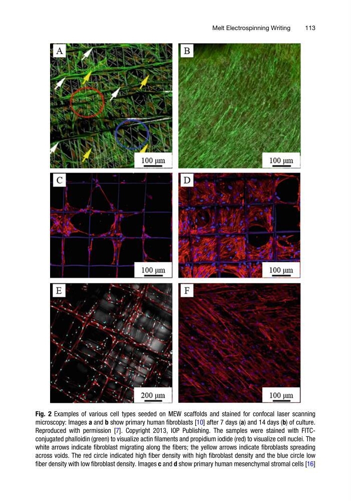
Fig. 2 Examples of various cell types seeded on MEW scaffolds and stained for confocal laser scanning
microscopy: Images a and b show primary human fibroblasts [10] after 7 days (a) and 14 days (b) of culture.
Reproduced with permission [7]. Copyright 2013, IOP Publishing. The samples were stained with FITCconjugated phalloidin (green) to visualize actin filaments and propidium iodide (red) to visualize cell nuclei. The
white arrows indicate fibroblast migrating along the fibers; the yellow arrows indicate fibroblasts spreading
across voids. The red circle indicated high fiber density with high fibroblast density and the blue circle low
fiber density with low fibroblast density. Images c and d show primary human mesenchymal stromal cells [16]
Melt Electrospinning Writing
113
microscopy: Images a and b show primary human fibroblasts [10] after 7 days (a) and 14 days (b) of culture.
Reproduced with permission [7]. Copyright 2013, IOP Publishing. The samples were stained with FITCconjugated phalloidin (green) to visualize actin filaments and propidium iodide (red) to visualize cell nuclei. The
white arrows indicate fibroblast migrating along the fibers; the yellow arrows indicate fibroblasts spreading
across voids. The red circle indicated high fiber density with high fibroblast density and the blue circle low
fiber density with low fibroblast density. Images c and d show primary human mesenchymal stromal cells [16]
Melt Electrospinning Writing
113