

Nanostructured Electrolytic Composites Based on Cobalt Alloys …
741
are equal to 0.1 and 0.2, accordingly, and are one order of magnitude higher than
those for Co–Mo–W coatings.
The Fe–Co–Mo coatings regardless of the deposition mode are characterized
by a more developed surface compared to the Co–Mo–W and Co–Mo–Zr alloys.
The AFM and SEM analyses show the Fe–Co–Mo coatings are micro-globular;
and agglomerates on the surface of 1–2 μm are formed from smaller grains (Fig. 1f,
g; Table 1).
The morphology and relief of Co–W–Zr coatings are similar to those of Co–Mo–
W coatings (Table 1, specimens 6, 7), however, the tungsten content reduces in the
alloys obtained for the same electrolysis conditions (Table 1, specimens 1 and 2).
It should be noted the zirconium percentage in Co–W–Zr is also lower in comparison to that in Co–Mo–Zr coatings deposited under the same conditions (Table 1,
specimens 3 and 5). A decrease in the size of associates (agglomerates) and unavailability of the cracks in the coatings can be related to the influence of tungsten on the
electro-crystallization process. An increase in the current density contributes to the
enrichment of the coating with tungsten, though the content of zirconium reduces to
1.78 wt% (Table 1, specimens 6 and 7).
Nevertheless, as it was shown in [21, 30–32] the relief and a degree of the surface
development of all the coatings mentioned above are favorable for the catalytic
processes that occur not only through the adsorption stage but are also realized in
the diffusive mode.
Alongside with the formation of branched globular surface the phase composition
of coatings can turn out to be an important factor that has an effect on their properties,
and in particular on the catalytic activity because it predetermines the distribution of
active accepting centers on the surface.
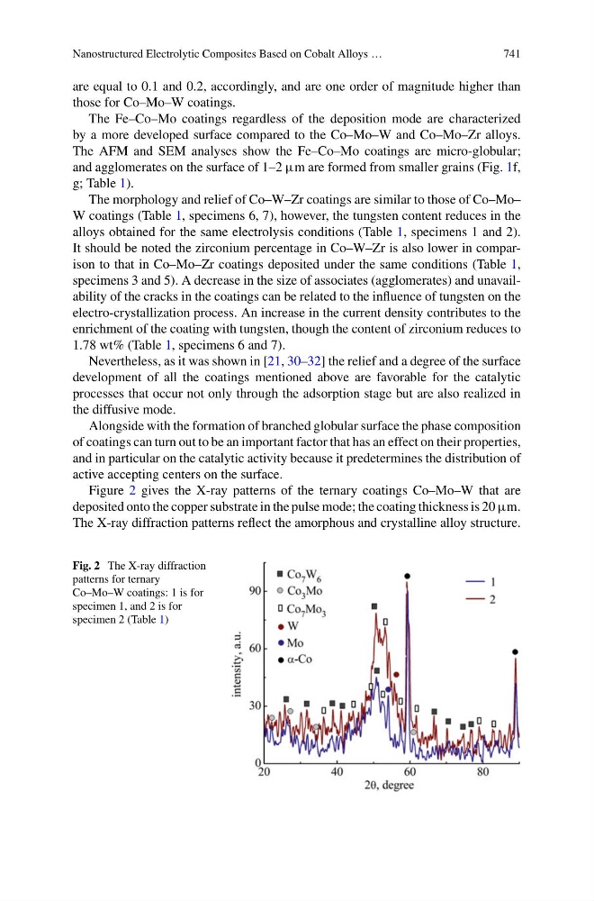
Figure 2 gives the X-ray patterns of the ternary coatings Co–Mo–W that are
deposited onto the copper substrate in the pulse mode; the coating thickness is 20 μm.
The X-ray diffraction patterns reflect the amorphous and crystalline alloy structure.
Fig. 2 The X-ray diffraction
patterns for ternary
Co–Mo–W coatings: 1 is for
specimen 1, and 2 is for
specimen 2 (Table 1)