

280
P. Raczy´ nski et al.
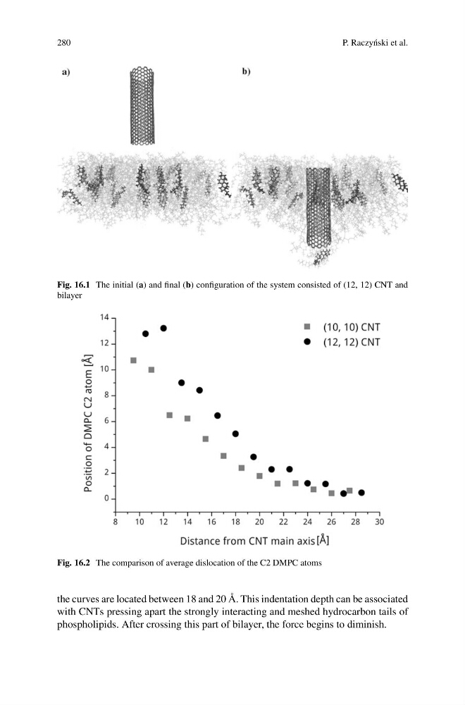
Fig. 16.1 The initial (a) and final (b) configuration of the system consisted of (12, 12) CNT and
bilayer
Fig. 16.2 The comparison of average dislocation of the C2 DMPC atoms
the curves are located between 18 and 20 Å. This indentation depth can be associated
with CNTs pressing apart the strongly interacting and meshed hydrocarbon tails of
phospholipids. After crossing this part of bilayer, the force begins to diminish.
P. Raczy´ nski et al.
Fig. 16.1 The initial (a) and final (b) configuration of the system consisted of (12, 12) CNT and
bilayer
Fig. 16.2 The comparison of average dislocation of the C2 DMPC atoms
the curves are located between 18 and 20 Å. This indentation depth can be associated
with CNTs pressing apart the strongly interacting and meshed hydrocarbon tails of
phospholipids. After crossing this part of bilayer, the force begins to diminish.