

213
Bowel Polyp Detection in Capsule Endoscopy Images
and Lu 2003). Because it is very challenging to obtain an accurate and clear
contour of polyps in CE images due to the complex background, in this chapter we turn to the region-based shape descriptor.
Taking into account the specific imaging circumstances required for CE
images mentioned previously, we need a shape feature that is invariant
to rotation, scale, and translation. Fortunately, Zernike moments satisfy
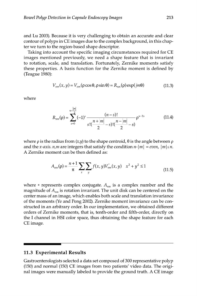
these properties. A basis function for the Zernike moment is defined by
(Teague 1980):
V nm (x, y) = V nm (ρcos θ, ρ sin θ) = R nm ( ρ)exp( jmθ)
(11.3)
where
m
n
−
( )
ρ = ∑
−
2
(−1 )
s
(n s )!
R nm
ρ
n −2s
(11.4)
n m
+
n m
−
s=0
s !(
− s)!(
− s s)
2
2
where ρ is the radius from (x,y) to the shape centroid, θ is the angle between ρ
and the x-axis. n,m are integers that satisfy the condition n-|m| = even, |m|≤ n.
A Zernike moment can be then defined as:
n + 1
A nm ( )
ρ =
∑ ∑ f x
( , y)V
*
nm (x, y) x
2
+ y
2
≤ 1
π
(11.5)
x
y
where * represents complex conjugate. A nm is a complex number and the
magnitude of A nm is rotation invariant. The unit disk can be centered on the
center mass of an image, which enables both scale and translation invariance
of the moments (Ye and Peng 2002). Zernike moment invariance can be constructed in an arbitrary order. In our implementation, we obtained different
orders of Zernike moments, that is, tenth-order and fifth-order, directly on
the I channel in HSI color space, thus obtaining the shape feature for each
CE image.
11.3 Experimental Results
Gastroenterologists selected a data set composed of 300 representative polyp
(150) and normal (150) CE images from two patients’ video data. The original images were manually labeled to provide the ground truth. A CE image
Bowel Polyp Detection in Capsule Endoscopy Images
and Lu 2003). Because it is very challenging to obtain an accurate and clear
contour of polyps in CE images due to the complex background, in this chapter we turn to the region-based shape descriptor.
Taking into account the specific imaging circumstances required for CE
images mentioned previously, we need a shape feature that is invariant
to rotation, scale, and translation. Fortunately, Zernike moments satisfy
these properties. A basis function for the Zernike moment is defined by
(Teague 1980):
V nm (x, y) = V nm (ρcos θ, ρ sin θ) = R nm ( ρ)exp( jmθ)
(11.3)
where
m
n
−
( )
ρ = ∑
−
2
(−1 )
s
(n s )!
R nm
ρ
n −2s
(11.4)
n m
+
n m
−
s=0
s !(
− s)!(
− s s)
2
2
where ρ is the radius from (x,y) to the shape centroid, θ is the angle between ρ
and the x-axis. n,m are integers that satisfy the condition n-|m| = even, |m|≤ n.
A Zernike moment can be then defined as:
n + 1
A nm ( )
ρ =
∑ ∑ f x
( , y)V
*
nm (x, y) x
2
+ y
2
≤ 1
π
(11.5)
x
y
where * represents complex conjugate. A nm is a complex number and the
magnitude of A nm is rotation invariant. The unit disk can be centered on the
center mass of an image, which enables both scale and translation invariance
of the moments (Ye and Peng 2002). Zernike moment invariance can be constructed in an arbitrary order. In our implementation, we obtained different
orders of Zernike moments, that is, tenth-order and fifth-order, directly on
the I channel in HSI color space, thus obtaining the shape feature for each
CE image.
11.3 Experimental Results
Gastroenterologists selected a data set composed of 300 representative polyp
(150) and normal (150) CE images from two patients’ video data. The original images were manually labeled to provide the ground truth. A CE image