

180
Biologically Inspired Robotics
Cantilever
Nanomanipulator
Tip
Cell
Aluminum substrate
Sharp tip
Flat tip
3 μm
3 μm
a
b
c
d
10 μm
δ
I
F
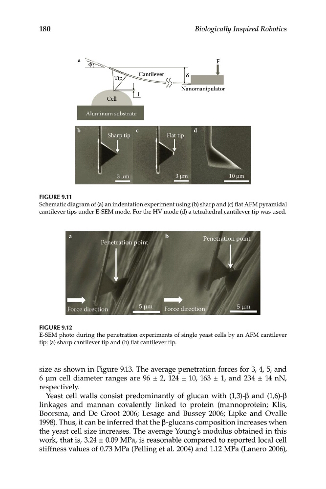
FIGURE 9.11
Schematic diagram of (a) an indentation experiment using (b) sharp and (c) flat AFM pyramidal
cantilever tips under E-SEM mode. For the HV mode (d) a tetrahedral cantilever tip was used.
a
b
Penetration point
Penetration point
5 μm
5 μm
Force direction
Force direction
FIGURE 9.12
E-SEM photo during the penetration experiments of single yeast cells by an AFM cantilever
tip: (a) sharp cantilever tip and (b) flat cantilever tip.
size as shown in Figure 9.13. The average penetration forces for 3, 4, 5, and
6 μm cell diameter ranges are 96 ± 2, 124 ± 10, 163 ± 1, and 234 ± 14 nN,
respectively.
Yeast cell walls consist predominantly of glucan with (1,3)-β and (1,6)-β
linkages and mannan covalently linked to protein (mannoprotein; Klis,
Boorsma, and De Groot 2006; Lesage and Bussey 2006; Lipke and Ovalle
1998). Thus, it can be inferred that the β-glucans composition increases when
the yeast cell size increases. The average Young’s modulus obtained in this
work, that is, 3.24 ± 0.09 MPa, is reasonable compared to reported local cell
stiffness values of 0.73 MPa (Pelling et al. 2004) and 1.12 MPa (Lanero 2006),
Biologically Inspired Robotics
Cantilever
Nanomanipulator
Tip
Cell
Aluminum substrate
Sharp tip
Flat tip
3 μm
3 μm
a
b
c
d
10 μm
δ
I
F
FIGURE 9.11
Schematic diagram of (a) an indentation experiment using (b) sharp and (c) flat AFM pyramidal
cantilever tips under E-SEM mode. For the HV mode (d) a tetrahedral cantilever tip was used.
a
b
Penetration point
Penetration point
5 μm
5 μm
Force direction
Force direction
FIGURE 9.12
E-SEM photo during the penetration experiments of single yeast cells by an AFM cantilever
tip: (a) sharp cantilever tip and (b) flat cantilever tip.
size as shown in Figure 9.13. The average penetration forces for 3, 4, 5, and
6 μm cell diameter ranges are 96 ± 2, 124 ± 10, 163 ± 1, and 234 ± 14 nN,
respectively.
Yeast cell walls consist predominantly of glucan with (1,3)-β and (1,6)-β
linkages and mannan covalently linked to protein (mannoprotein; Klis,
Boorsma, and De Groot 2006; Lesage and Bussey 2006; Lipke and Ovalle
1998). Thus, it can be inferred that the β-glucans composition increases when
the yeast cell size increases. The average Young’s modulus obtained in this
work, that is, 3.24 ± 0.09 MPa, is reasonable compared to reported local cell
stiffness values of 0.73 MPa (Pelling et al. 2004) and 1.12 MPa (Lanero 2006),