

130
Biologically Inspired Robotics
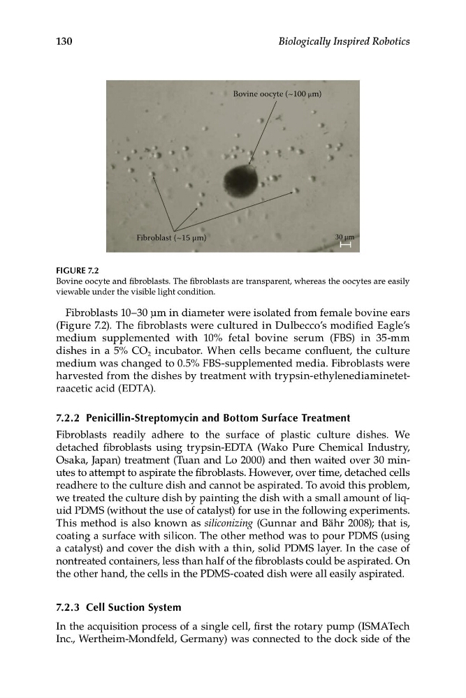
Bovine oocyte (~100 μm)
Fibroblast (~15 μm)
30 μm
FIGURE 7.2
Bovine oocyte and fibroblasts. The fibroblasts are transparent, whereas the oocytes are easily
viewable under the visible light condition.
Fibroblasts 10–30 μm in diameter were isolated from female bovine ears
(Figure 7.2). The fibroblasts were cultured in Dulbecco’s modified Eagle’s
medium supplemented with 10% fetal bovine serum (FBS) in 35-mm
dishes in a 5% CO 2 incubator. When cells became confluent, the culture
medium was changed to 0.5% FBS-supplemented media. Fibroblasts were
harvested from the dishes by treatment with trypsin-ethylenediaminetetraacetic acid (EDTA).
7.2.2 Penicillin-Streptomycin and Bottom Surface Treatment
Fibroblasts readily adhere to the surface of plastic culture dishes. We
detached fibroblasts using trypsin-EDTA (Wako Pure Chemical Industry,
Osaka, Japan) treatment (Tuan and Lo 2000) and then waited over 30 minutes to attempt to aspirate the fibroblasts. However, over time, detached cells
readhere to the culture dish and cannot be aspirated. To avoid this problem,
we treated the culture dish by painting the dish with a small amount of liquid PDMS (without the use of catalyst) for use in the following experiments.
This method is also known as siliconizing (Gunnar and Bähr 2008); that is,
coating a surface with silicon. The other method was to pour PDMS (using
a catalyst) and cover the dish with a thin, solid PDMS layer. In the case of
nontreated containers, less than half of the fibroblasts could be aspirated. On
the other hand, the cells in the PDMS-coated dish were all easily aspirated.
7.2.3 Cell Suction System
In the acquisition process of a single cell, first the rotary pump (ISMATech
Inc., Wertheim-Mondfeld, Germany) was connected to the dock side of the
Biologically Inspired Robotics
Bovine oocyte (~100 μm)
Fibroblast (~15 μm)
30 μm
FIGURE 7.2
Bovine oocyte and fibroblasts. The fibroblasts are transparent, whereas the oocytes are easily
viewable under the visible light condition.
Fibroblasts 10–30 μm in diameter were isolated from female bovine ears
(Figure 7.2). The fibroblasts were cultured in Dulbecco’s modified Eagle’s
medium supplemented with 10% fetal bovine serum (FBS) in 35-mm
dishes in a 5% CO 2 incubator. When cells became confluent, the culture
medium was changed to 0.5% FBS-supplemented media. Fibroblasts were
harvested from the dishes by treatment with trypsin-ethylenediaminetetraacetic acid (EDTA).
7.2.2 Penicillin-Streptomycin and Bottom Surface Treatment
Fibroblasts readily adhere to the surface of plastic culture dishes. We
detached fibroblasts using trypsin-EDTA (Wako Pure Chemical Industry,
Osaka, Japan) treatment (Tuan and Lo 2000) and then waited over 30 minutes to attempt to aspirate the fibroblasts. However, over time, detached cells
readhere to the culture dish and cannot be aspirated. To avoid this problem,
we treated the culture dish by painting the dish with a small amount of liquid PDMS (without the use of catalyst) for use in the following experiments.
This method is also known as siliconizing (Gunnar and Bähr 2008); that is,
coating a surface with silicon. The other method was to pour PDMS (using
a catalyst) and cover the dish with a thin, solid PDMS layer. In the case of
nontreated containers, less than half of the fibroblasts could be aspirated. On
the other hand, the cells in the PDMS-coated dish were all easily aspirated.
7.2.3 Cell Suction System
In the acquisition process of a single cell, first the rotary pump (ISMATech
Inc., Wertheim-Mondfeld, Germany) was connected to the dock side of the