

Compact vision
Next module
system
Supply
Syringe pump
Valves
PDMS microfluidic
chip
Docking area
Compact
Supply
Suction
vision system
Suction
Monitor
Container
Rotary pump
XY-stage
XY-stage
129
Automatic Single-Cell Transfer Module
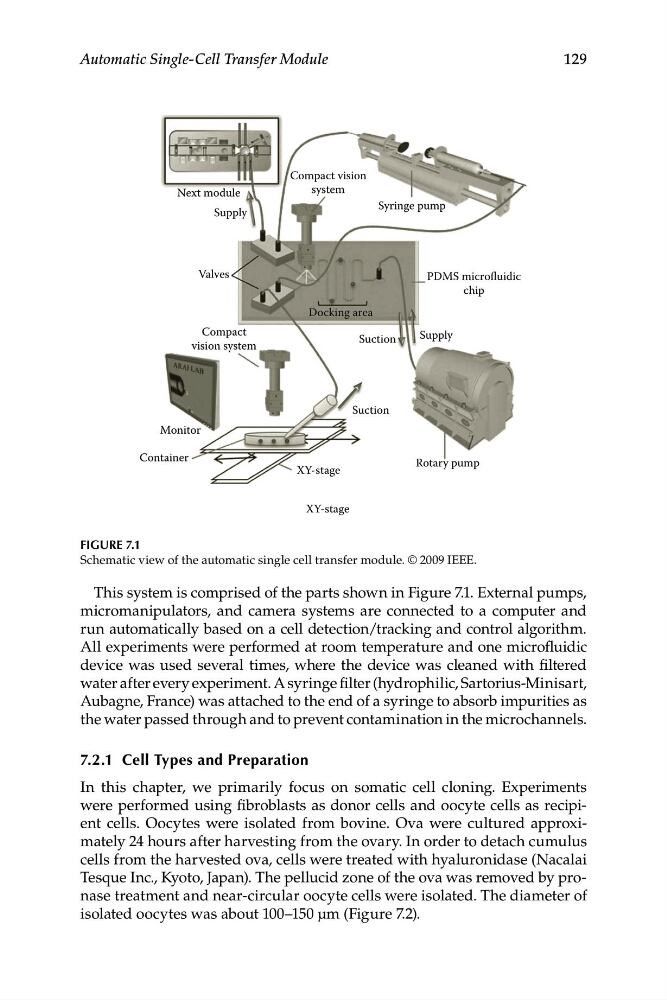
FIGURE 7.1
Schematic view of the automatic single cell transfer module. © 2009 IEEE.
This system is comprised of the parts shown in Figure 7.1. External pumps,
micromanipulators, and camera systems are connected to a computer and
run automatically based on a cell detection/tracking and control algorithm.
All experiments were performed at room temperature and one microfluidic
device was used several times, where the device was cleaned with filtered
water after every experiment. A syringe filter (hydrophilic, Sartorius-Minisart,
Aubagne, France) was attached to the end of a syringe to absorb impurities as
the water passed through and to prevent contamination in the microchannels.
7.2.1 Cell Types and Preparation
In this chapter, we primarily focus on somatic cell cloning. Experiments
were performed using fibroblasts as donor cells and oocyte cells as recipient cells. Oocytes were isolated from bovine. Ova were cultured approximately 24 hours after harvesting from the ovary. In order to detach cumulus
cells from the harvested ova, cells were treated with hyaluronidase (Nacalai
Tesque Inc., Kyoto, Japan). The pellucid zone of the ova was removed by pronase treatment and near-circular oocyte cells were isolated. The diameter of
isolated oocytes was about 100–150 μm (Figure 7.2).
Next module
system
Supply
Syringe pump
Valves
PDMS microfluidic
chip
Docking area
Compact
Supply
Suction
vision system
Suction
Monitor
Container
Rotary pump
XY-stage
XY-stage
129
Automatic Single-Cell Transfer Module
FIGURE 7.1
Schematic view of the automatic single cell transfer module. © 2009 IEEE.
This system is comprised of the parts shown in Figure 7.1. External pumps,
micromanipulators, and camera systems are connected to a computer and
run automatically based on a cell detection/tracking and control algorithm.
All experiments were performed at room temperature and one microfluidic
device was used several times, where the device was cleaned with filtered
water after every experiment. A syringe filter (hydrophilic, Sartorius-Minisart,
Aubagne, France) was attached to the end of a syringe to absorb impurities as
the water passed through and to prevent contamination in the microchannels.
7.2.1 Cell Types and Preparation
In this chapter, we primarily focus on somatic cell cloning. Experiments
were performed using fibroblasts as donor cells and oocyte cells as recipient cells. Oocytes were isolated from bovine. Ova were cultured approximately 24 hours after harvesting from the ovary. In order to detach cumulus
cells from the harvested ova, cells were treated with hyaluronidase (Nacalai
Tesque Inc., Kyoto, Japan). The pellucid zone of the ova was removed by pronase treatment and near-circular oocyte cells were isolated. The diameter of
isolated oocytes was about 100–150 μm (Figure 7.2).