

4.1 Introduction
71
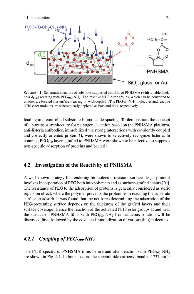
Scheme 4.1 Schematic structure of substrate-supported thin film of PNHSMA (with tunable thickness d film ) reacting with PEG 500 -NH 2 . The reactive NHS ester groups, which can be converted to
amides, are located in a surface-near region with depth d z . The PEG 500 -NH 2 molecules and reactive
NHS ester moieties are schematically depicted as bars and dots, respectively
loading and controlled substrate-biomolecule spacing. To demonstrate the concept
of a biosensor architecture for pathogen detection based on the PNHSMA platform,
anti-listeria antibodies, immobilized via strong interactions with covalently coupled
and correctly oriented protein G, were shown to selectively recognize listeria. In
contrast, PEG 500 layers grafted to PNHSMA were shown to be effective to suppress
non-specific adsorption of proteins and bacteria.
4.2 Investigation of the Reactivity of PNHSMA
A well-known strategy for rendering biomolecule-resistant surfaces (e.g., protein)
involves incorporation of PEG both into polymers and as surface-grafted chains [20].
The resistance of PEG to the adsorption of proteins is generally considered as steric
repulsion effect, where the polymer prevents the protein from reaching the substrate
surface to adsorb. It was found that the net force determining the adsorption of the
PEG-presenting surface depends on the thickness of the grafted layers and their
surface coverage. Hence the reaction of the activated NHS ester groups at and near
the surface of PNHSMA films with PEG 500 -NH 2 from aqueous solution will be
discussed first, followed by the covalent immobilization of various (bio)molecules.
4.2.1 Coupling of PEG 500 -NH 2
The FTIR spectra of PNHSMA films before and after reaction with PEG 500 -NH 2
are shown in Fig. 4.1. In both spectra, the succinimide carbonyl band at 1737 cm
−1
71
Scheme 4.1 Schematic structure of substrate-supported thin film of PNHSMA (with tunable thickness d film ) reacting with PEG 500 -NH 2 . The reactive NHS ester groups, which can be converted to
amides, are located in a surface-near region with depth d z . The PEG 500 -NH 2 molecules and reactive
NHS ester moieties are schematically depicted as bars and dots, respectively
loading and controlled substrate-biomolecule spacing. To demonstrate the concept
of a biosensor architecture for pathogen detection based on the PNHSMA platform,
anti-listeria antibodies, immobilized via strong interactions with covalently coupled
and correctly oriented protein G, were shown to selectively recognize listeria. In
contrast, PEG 500 layers grafted to PNHSMA were shown to be effective to suppress
non-specific adsorption of proteins and bacteria.
4.2 Investigation of the Reactivity of PNHSMA
A well-known strategy for rendering biomolecule-resistant surfaces (e.g., protein)
involves incorporation of PEG both into polymers and as surface-grafted chains [20].
The resistance of PEG to the adsorption of proteins is generally considered as steric
repulsion effect, where the polymer prevents the protein from reaching the substrate
surface to adsorb. It was found that the net force determining the adsorption of the
PEG-presenting surface depends on the thickness of the grafted layers and their
surface coverage. Hence the reaction of the activated NHS ester groups at and near
the surface of PNHSMA films with PEG 500 -NH 2 from aqueous solution will be
discussed first, followed by the covalent immobilization of various (bio)molecules.
4.2.1 Coupling of PEG 500 -NH 2
The FTIR spectra of PNHSMA films before and after reaction with PEG 500 -NH 2
are shown in Fig. 4.1. In both spectra, the succinimide carbonyl band at 1737 cm
−1