

48
3 Confinement Effects on the Reactivity in Ultrathin …
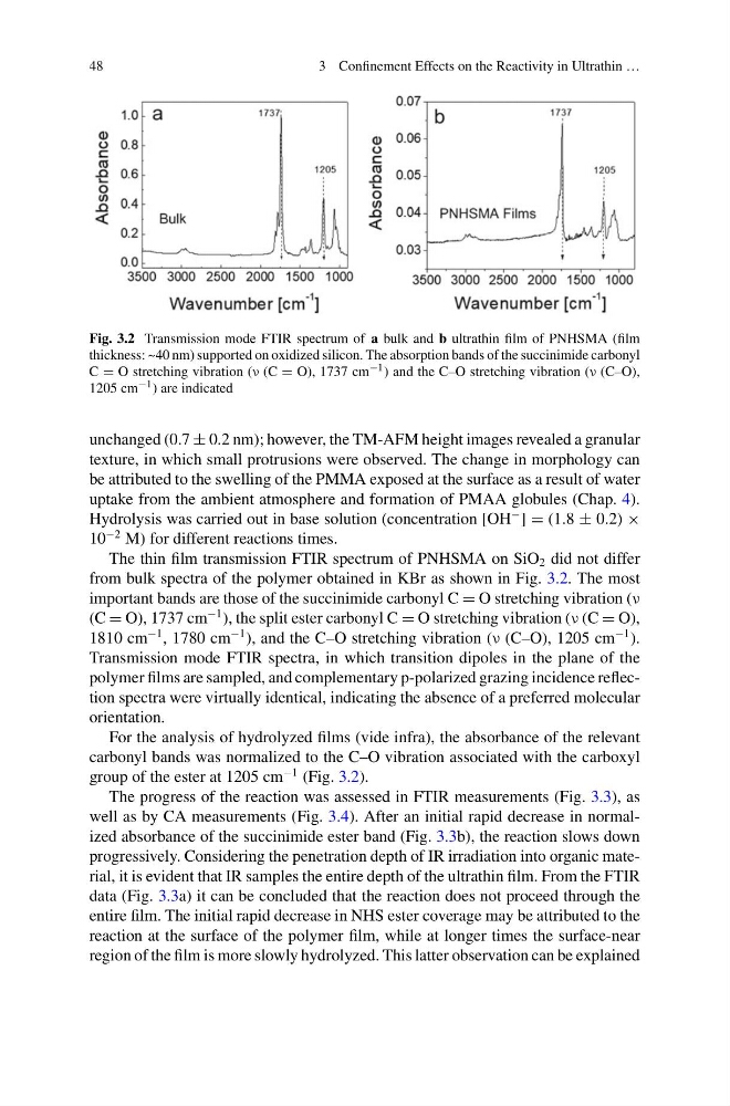
Fig. 3.2 Transmission mode FTIR spectrum of a bulk and b ultrathin film of PNHSMA (film
thickness: ~40 nm) supported on oxidized silicon. The absorption bands of the succinimide carbonyl
C = O stretching vibration (ν (C = O), 1737 cm −1 ) and the C–O stretching vibration (ν (C–O),
1205 cm −1 ) are indicated
unchanged (0.7 ± 0.2 nm); however, the TM-AFM height images revealed a granular
texture, in which small protrusions were observed. The change in morphology can
be attributed to the swelling of the PMMA exposed at the surface as a result of water
uptake from the ambient atmosphere and formation of PMAA globules (Chap. 4).
Hydrolysis was carried out in base solution (concentration [OH
− ] = (1.8 ± 0.2) ×
10
−2 M) for different reactions times.
The thin film transmission FTIR spectrum of PNHSMA on SiO 2 did not differ
from bulk spectra of the polymer obtained in KBr as shown in Fig. 3.2. The most
important bands are those of the succinimide carbonyl C = O stretching vibration (ν
(C = O), 1737 cm
−1 ), the split ester carbonyl C = O stretching vibration (ν (C = O),
1810 cm
−1 , 1780 cm
−1 ), and the C–O stretching vibration (ν (C–O), 1205 cm
−1 ).
Transmission mode FTIR spectra, in which transition dipoles in the plane of the
polymer films are sampled, and complementary p-polarized grazing incidence reflection spectra were virtually identical, indicating the absence of a preferred molecular
orientation.
For the analysis of hydrolyzed films (vide infra), the absorbance of the relevant
carbonyl bands was normalized to the C–O vibration associated with the carboxyl
group of the ester at 1205 cm
−1 (Fig. 3.2).
The progress of the reaction was assessed in FTIR measurements (Fig. 3.3), as
well as by CA measurements (Fig. 3.4). After an initial rapid decrease in normalized absorbance of the succinimide ester band (Fig. 3.3b), the reaction slows down
progressively. Considering the penetration depth of IR irradiation into organic material, it is evident that IR samples the entire depth of the ultrathin film. From the FTIR
data (Fig. 3.3a) it can be concluded that the reaction does not proceed through the
entire film. The initial rapid decrease in NHS ester coverage may be attributed to the
reaction at the surface of the polymer film, while at longer times the surface-near
region of the film is more slowly hydrolyzed. This latter observation can be explained
3 Confinement Effects on the Reactivity in Ultrathin …
Fig. 3.2 Transmission mode FTIR spectrum of a bulk and b ultrathin film of PNHSMA (film
thickness: ~40 nm) supported on oxidized silicon. The absorption bands of the succinimide carbonyl
C = O stretching vibration (ν (C = O), 1737 cm −1 ) and the C–O stretching vibration (ν (C–O),
1205 cm −1 ) are indicated
unchanged (0.7 ± 0.2 nm); however, the TM-AFM height images revealed a granular
texture, in which small protrusions were observed. The change in morphology can
be attributed to the swelling of the PMMA exposed at the surface as a result of water
uptake from the ambient atmosphere and formation of PMAA globules (Chap. 4).
Hydrolysis was carried out in base solution (concentration [OH
− ] = (1.8 ± 0.2) ×
10
−2 M) for different reactions times.
The thin film transmission FTIR spectrum of PNHSMA on SiO 2 did not differ
from bulk spectra of the polymer obtained in KBr as shown in Fig. 3.2. The most
important bands are those of the succinimide carbonyl C = O stretching vibration (ν
(C = O), 1737 cm
−1 ), the split ester carbonyl C = O stretching vibration (ν (C = O),
1810 cm
−1 , 1780 cm
−1 ), and the C–O stretching vibration (ν (C–O), 1205 cm
−1 ).
Transmission mode FTIR spectra, in which transition dipoles in the plane of the
polymer films are sampled, and complementary p-polarized grazing incidence reflection spectra were virtually identical, indicating the absence of a preferred molecular
orientation.
For the analysis of hydrolyzed films (vide infra), the absorbance of the relevant
carbonyl bands was normalized to the C–O vibration associated with the carboxyl
group of the ester at 1205 cm
−1 (Fig. 3.2).
The progress of the reaction was assessed in FTIR measurements (Fig. 3.3), as
well as by CA measurements (Fig. 3.4). After an initial rapid decrease in normalized absorbance of the succinimide ester band (Fig. 3.3b), the reaction slows down
progressively. Considering the penetration depth of IR irradiation into organic material, it is evident that IR samples the entire depth of the ultrathin film. From the FTIR
data (Fig. 3.3a) it can be concluded that the reaction does not proceed through the
entire film. The initial rapid decrease in NHS ester coverage may be attributed to the
reaction at the surface of the polymer film, while at longer times the surface-near
region of the film is more slowly hydrolyzed. This latter observation can be explained