

32
2 Surface Reactions and Fabrication of Bioreactive Platforms …
the hydrophobic regions of the SAM. By using μCP, biotin was also transferred onto
mixed SAMs of alkanethiolates on gold, which were made from thiols presenting
terminal tri(ethylene glycol) groups and terminal hexa(ethylene glycol) groups. The
formation of patterned SAMs presenting biotin ligands was detected by fluorescence
microscopy of substrates that were incubated with a solution of fluorescently labeled
antibiotin antibody.
Patterning biotin and streptavidin on a polymer surface by physical adsorption has
been studied using reactive μCP by Hyun et al. [50]. They demonstrate that polyethylene, polystyrene, poly(methyl methacrylate), and poly(ethylene terephthalate) films
have been successfully patterned using biotin. By exploiting molecular recognition
between biotin and streptavidin, streptavidin was adsorbed on the micropatterned
biotin surfaces, as detected by fluorescence microscopy.
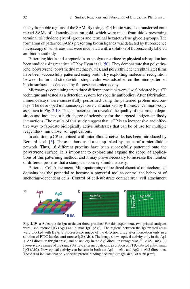
Microarrays containing up to three different proteins were also fabricated by μCP
technique and tested as a detection system for specific antibodies. After fabrication,
immunoassays were successfully performed using the patterned protein microarrays. The developed immunoassays were characterized by fluorescence microscopy
as shown in Fig. 2.19. The characterization revealed the quality of the protein deposition and indicated a high degree of selectivity for the targeted antigen–antibody
interactions. The results of this study suggest that μCP is an inexpensive and effective way to fabricate biologically active substrates that can be of use for multiple
reagentless immunosensor applications.
In addition, μCP combined with microfluidic networks has been introduced by
Bernard et al. [5]. These authors used a stamp inked by means of a microfluidic
network. Thus, 16 different proteins have been successfully patterned onto the
polystyrene surface. It is important to explore and expand the scope of applications of this patterning method, and it may prove necessary to increase the number
of different proteins that a stamp can convey simultaneously.
Patterned Cell Attachment: Micropatterning of localized chemical or biochemical
domains has the potential to become a powerful tool to control the behavior of
anchorage-dependent cells. Control of cell-substrate contact area, cell attachment
Fig. 2.19 a Substrate design to detect three proteins. For this experiment, two printed antigens
were used, mouse IgG (Ag1) and human IgG (Ag2). The regions between the IgGprinted areas
were blocked with BSA. b Fluorescence image of the detection array after incubation only in a
solution of FTIC-labeled anti-mouse IgG (Ab1). The image shows optical activity only in the Ag1
+ Ab1 direction (bright areas) and no activity in the Ag2 direction (image size, 30 × 45 μm 2 ). (c)
Fluorescence image of the same substrate after incubation in a solution of FTIC-labeled anti-human
IgG (Ab2). Now optical activity can be seen in both the Ag1 + Ab1 and Ag2 + Ab2 directions.
These data indicate that only specific protein binding occurred (image size, 30 × 56 μm 2 )
2 Surface Reactions and Fabrication of Bioreactive Platforms …
the hydrophobic regions of the SAM. By using μCP, biotin was also transferred onto
mixed SAMs of alkanethiolates on gold, which were made from thiols presenting
terminal tri(ethylene glycol) groups and terminal hexa(ethylene glycol) groups. The
formation of patterned SAMs presenting biotin ligands was detected by fluorescence
microscopy of substrates that were incubated with a solution of fluorescently labeled
antibiotin antibody.
Patterning biotin and streptavidin on a polymer surface by physical adsorption has
been studied using reactive μCP by Hyun et al. [50]. They demonstrate that polyethylene, polystyrene, poly(methyl methacrylate), and poly(ethylene terephthalate) films
have been successfully patterned using biotin. By exploiting molecular recognition
between biotin and streptavidin, streptavidin was adsorbed on the micropatterned
biotin surfaces, as detected by fluorescence microscopy.
Microarrays containing up to three different proteins were also fabricated by μCP
technique and tested as a detection system for specific antibodies. After fabrication,
immunoassays were successfully performed using the patterned protein microarrays. The developed immunoassays were characterized by fluorescence microscopy
as shown in Fig. 2.19. The characterization revealed the quality of the protein deposition and indicated a high degree of selectivity for the targeted antigen–antibody
interactions. The results of this study suggest that μCP is an inexpensive and effective way to fabricate biologically active substrates that can be of use for multiple
reagentless immunosensor applications.
In addition, μCP combined with microfluidic networks has been introduced by
Bernard et al. [5]. These authors used a stamp inked by means of a microfluidic
network. Thus, 16 different proteins have been successfully patterned onto the
polystyrene surface. It is important to explore and expand the scope of applications of this patterning method, and it may prove necessary to increase the number
of different proteins that a stamp can convey simultaneously.
Patterned Cell Attachment: Micropatterning of localized chemical or biochemical
domains has the potential to become a powerful tool to control the behavior of
anchorage-dependent cells. Control of cell-substrate contact area, cell attachment
Fig. 2.19 a Substrate design to detect three proteins. For this experiment, two printed antigens
were used, mouse IgG (Ag1) and human IgG (Ag2). The regions between the IgGprinted areas
were blocked with BSA. b Fluorescence image of the detection array after incubation only in a
solution of FTIC-labeled anti-mouse IgG (Ab1). The image shows optical activity only in the Ag1
+ Ab1 direction (bright areas) and no activity in the Ag2 direction (image size, 30 × 45 μm 2 ). (c)
Fluorescence image of the same substrate after incubation in a solution of FTIC-labeled anti-human
IgG (Ab2). Now optical activity can be seen in both the Ag1 + Ab1 and Ag2 + Ab2 directions.
These data indicate that only specific protein binding occurred (image size, 30 × 56 μm 2 )