

9.4 Conclusion and Outlook
179
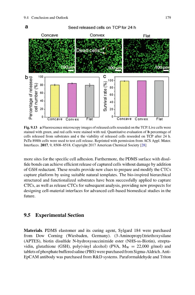
Fig. 9.13 a Fluorescence microscopy images of released cells reseeded on the TCP. Live cells were
stained with green, and red cells were stained with red. Quantitative evaluation of b percentage of
cells released from substrates and c the viability of released cells reseeded on TCP after 24 h.
PaTu 8988t cells were used to test cell release. Reprinted with permission from ACS Appl. Mater.
Interfaces. 2017, 9, 8508–8518. Copyright 2017 American Chemical Society [28]
more sites for the specific cell adhesion. Furthermore, the PDMS surface with disulfide bonds can achieve efficient release of captured cells without damage by addition
of GSH reductant. These results provide new clues to prepare and modify the CTCs
capture platform by using suitable natural templates. The bio-inspired hierarchical
structured and functionalized substrates have been successfully applied to capture
CTCs, as well as release CTCs for subsequent analysis, providing new prospects for
designing cell-material interfaces for advanced cell-based biomedical studies in the
future.
9.5 Experimental Section
Materials. PDMS elastomer and its curing agent, Sylgard 184 were purchased
from Dow Corning (Wiesbaden, Germany). (3-Aminopropyl)triethoxysilane
(APTES), biotin disulfide N-hydroxysuccinimide ester (NHS-ss-Biotin), streptavidin, glutathione (GSH), poly(vinyl alcohol) (PVA, M W = 22,000 g/mol) and
tablets of phosphate buffered saline (PBS) were purchased from Sigma-Aldrich. AntiEpCAM antibody was purchased from R&D systems. Paraformaldehyde and Triton
179
Fig. 9.13 a Fluorescence microscopy images of released cells reseeded on the TCP. Live cells were
stained with green, and red cells were stained with red. Quantitative evaluation of b percentage of
cells released from substrates and c the viability of released cells reseeded on TCP after 24 h.
PaTu 8988t cells were used to test cell release. Reprinted with permission from ACS Appl. Mater.
Interfaces. 2017, 9, 8508–8518. Copyright 2017 American Chemical Society [28]
more sites for the specific cell adhesion. Furthermore, the PDMS surface with disulfide bonds can achieve efficient release of captured cells without damage by addition
of GSH reductant. These results provide new clues to prepare and modify the CTCs
capture platform by using suitable natural templates. The bio-inspired hierarchical
structured and functionalized substrates have been successfully applied to capture
CTCs, as well as release CTCs for subsequent analysis, providing new prospects for
designing cell-material interfaces for advanced cell-based biomedical studies in the
future.
9.5 Experimental Section
Materials. PDMS elastomer and its curing agent, Sylgard 184 were purchased
from Dow Corning (Wiesbaden, Germany). (3-Aminopropyl)triethoxysilane
(APTES), biotin disulfide N-hydroxysuccinimide ester (NHS-ss-Biotin), streptavidin, glutathione (GSH), poly(vinyl alcohol) (PVA, M W = 22,000 g/mol) and
tablets of phosphate buffered saline (PBS) were purchased from Sigma-Aldrich. AntiEpCAM antibody was purchased from R&D systems. Paraformaldehyde and Triton