

9.2 Preparation of Rose Petal-Derived PDMS Surface Functionalized …
169
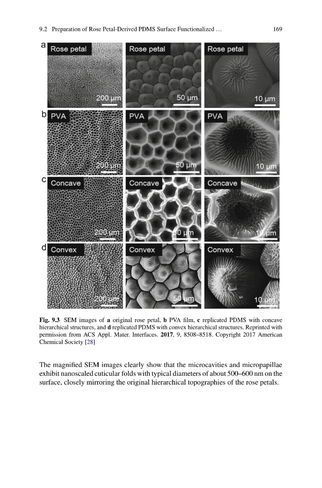
Fig. 9.3 SEM images of a original rose petal, b PVA film, c replicated PDMS with concave
hierarchical structures, and d replicated PDMS with convex hierarchical structures. Reprinted with
permission from ACS Appl. Mater. Interfaces. 2017, 9, 8508–8518. Copyright 2017 American
Chemical Society [28]
The magnified SEM images clearly show that the microcavities and micropapillae
exhibit nanoscaled cuticular folds with typical diameters of about 500–600 nm on the
surface, closely mirroring the original hierarchical topographies of the rose petals.
169
Fig. 9.3 SEM images of a original rose petal, b PVA film, c replicated PDMS with concave
hierarchical structures, and d replicated PDMS with convex hierarchical structures. Reprinted with
permission from ACS Appl. Mater. Interfaces. 2017, 9, 8508–8518. Copyright 2017 American
Chemical Society [28]
The magnified SEM images clearly show that the microcavities and micropapillae
exhibit nanoscaled cuticular folds with typical diameters of about 500–600 nm on the
surface, closely mirroring the original hierarchical topographies of the rose petals.